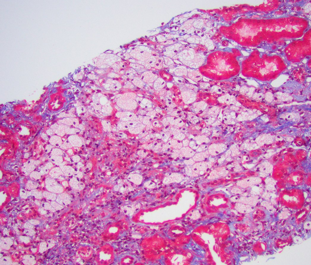
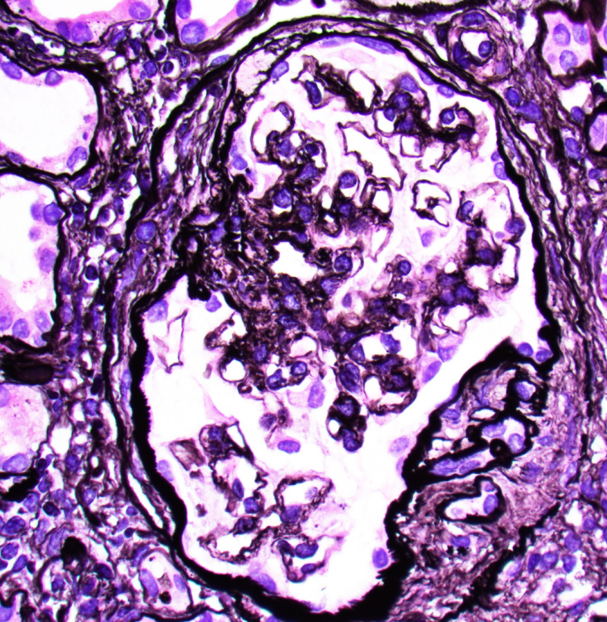
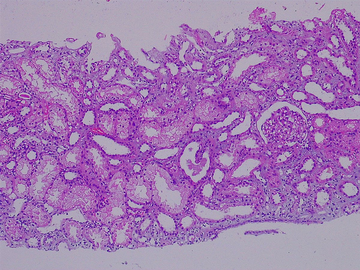
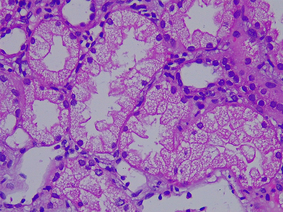
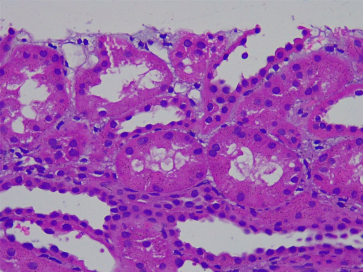
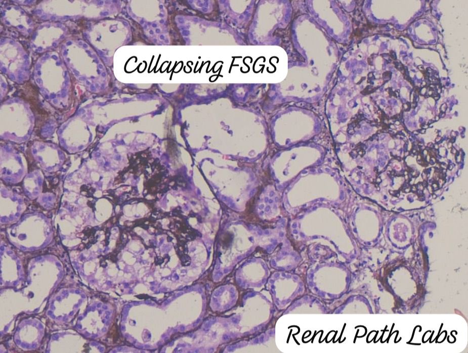
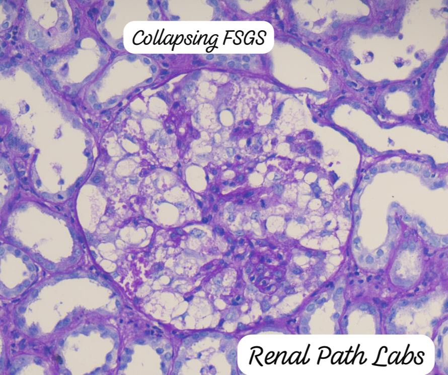
Renal Path Labs

934 posts

Renal Path Labs banner
Renal Path Labs

Renal Path Labs

@RenalPathLabs

Renal Pathologist and Literature enthusiast!! Lifelong learner. Tweets are my own!

Gurgaon, India Katılım Eylül 2013
1.2K Takip Edilen1.6K Takipçiler
Renal Path Labs retweetledi
Sam Albadri, M.D., M.Sc.
Sam Albadri, M.D., M.Sc.@sam_albadri·
📌 Renal Pathology Pearl : ✅Light Chain-Only PGNMID🔦 Light chain–only PGNMID (PMID: 32001067 ) shows monotypic light chain deposits (κ or λ) with absent heavy chains , a true light chain–only pattern. 💡 Key features: • IF: Isolated light chain restriction ± C3 co-staining (reflects alternative pathway activation, not primary C3 glomerulopathy). • LM: MPGN, endocapillary proliferative, or mesangial proliferative patterns. • EM: Granular, electron-dense deposits (non-organized). • Always evaluate for underlying B-cell/plasma cell clone. 📍 C3 positivity is common and due to complement activation, but not C3G — diagnosis rests on true monoclonal light chain restriction. @Renalpathsoc @MCFNephFellows @MayoClinicNeph @MayoClinicPath
English
1
14
35
1.7K
Renal Path Labs retweetledi
Sam Albadri, M.D., M.Sc.
Sam Albadri, M.D., M.Sc.@sam_albadri·
🧠🩺 Renal Pathology Pearl: Lysozyme (Lysosomal) Nephropathy 📌 Often seen in CMML / monocytic leukemias
📌 Causes tubular (non-albumin) proteinuria 🔬 Light Microscopy
• Predominant tubulointerstitial injury
• Proximal tubules with eosinophilic, granular/vacuolated cytoplasm
• Interstitial inflammation ± fibrosis
• Glomeruli largely unremarkable 🧪 Immunofluorescence
• Negative for IgG, IgA, IgM
• Negative kappa/lambda
• C3 absent or nonspecific
• (If performed: strong cytoplasmic lysozyme staining in proximal tubules) 🔍 Electron Microscopy
• Proximal tubular cells packed with enlarged lysosomes
• Lysosomes contain electron-dense granular ± crystalline material
• No immune-type deposits in glomeruli or TBMs 🧩 Key clue: High UPCR + low ACR → non-albumin proteinuria
🚫 Argues against AL amyloid, LCDD, PGNMID #RenalPath #NephPath #LysozymeNephropathy #KidneyBiopsy #MedEd @MayoClinicPath
Sam Albadri, M.D., M.Sc. tweet media
Română
2
34
73
3.2K
Sijie
Sijie@sijiezheng·
@ChangUCanSpare Maybe IgA is a “Pattern of injury “? Just like FSGS, MPGN
English
1
0
1
56
Renal Path Labs retweetledi
Arkana Laboratories
Arkana Laboratories@arkanalabs·
The light microscopic image depicts interstitial foam cells which, while non-specific and can be seen in longstanding proteinuria, are concerning for Alport Syndrome given the clinical scenario of hematuria and hearing loss. By electron microscopy, diagnostic findings would include glomerular basement membrane splitting, scalloping, multilamellation or "basket-weaving", intramembranous "bread crumbs", and abrupt transitions from areas of thin and thick GBMs. An Alport staining panel will classically show loss of staining for collagen IV alpha 5 in the majority of cases (~85%, X-linked inheritance). #renalpath #kidneypath #pathology #renal #pathX
Arkana Laboratories@arkanalabs

What is this finding? In a young male patient with hematuria and hearing loss, what would be your top diagnosis? #DiagnoseThis #pathology #renal #kidneypath #pathx #pathtwitter

English
1
9
26
2.3K
Renal Path Labs retweetledi
Jonathan Zuckerman MD PhD
Jonathan Zuckerman MD PhD@JZRenalPath·
Classic example of Alport spectrum disease in a pt with X-linked Alport syndrome. Lesions of segmental glomerulosclerosis, numerous interstitial foam cells, and atypical GBMs with lamina densa splitting, subepithelial scalloping, and thinning. #renalpath #pathtwitter #nephrology
Jonathan Zuckerman MD PhD tweet mediaJonathan Zuckerman MD PhD tweet mediaJonathan Zuckerman MD PhD tweet mediaJonathan Zuckerman MD PhD tweet media
English
0
23
60
2.2K
Renal Path Labs retweetledi
Anthony Chang, MD (張賀文)
Anthony Chang, MD (張賀文)@ChangUCanSpare·
Random reminder: hematuria in thin basement membrane nephropathy. Is this how it occurs? #renalpath
Anthony Chang, MD (張賀文)@ChangUCanSpare

@podocytes Feast your eyes on this figure (1 of 4 pgs) from 2001 KI study. Still can't wrap my head around how this actually happens. How does the #RBC go through the #podocyte like it's not even there? #SeeingIsBelievingOrIsIt ncbi.nlm.nih.gov/pubmed/11380808

English
4
24
72
0
Renal Path Labs retweetledi
Sam Albadri, M.D., M.Sc.
Sam Albadri, M.D., M.Sc.@sam_albadri·
Electron microscopy examination of the Kappa light chain cast nephropathy reveals atypical casts characterized by granular, electron-dense material and fibrillar texture deposits #renalpath
Sam Albadri, M.D., M.Sc. tweet mediaSam Albadri, M.D., M.Sc. tweet media
English
3
8
32
1.9K
Mahabub Alam
Mahabub Alam@docrobin52·
@RenalPathLabs @Renalpathsoc Don’t see kidney biopsy, but I’ll try. Silver stain showing vasculitis of a medium sized artery, granular casts and dense interstitial inflammation. 🤔
English
1
0
1
78