

Alexander Crichton
671 posts

@VascularABC
Vascular research fellow @Houston Methodist hospital, University of Birmingham PhD Student, OOPR West Midlands Vascular Resident, Father and Husband!


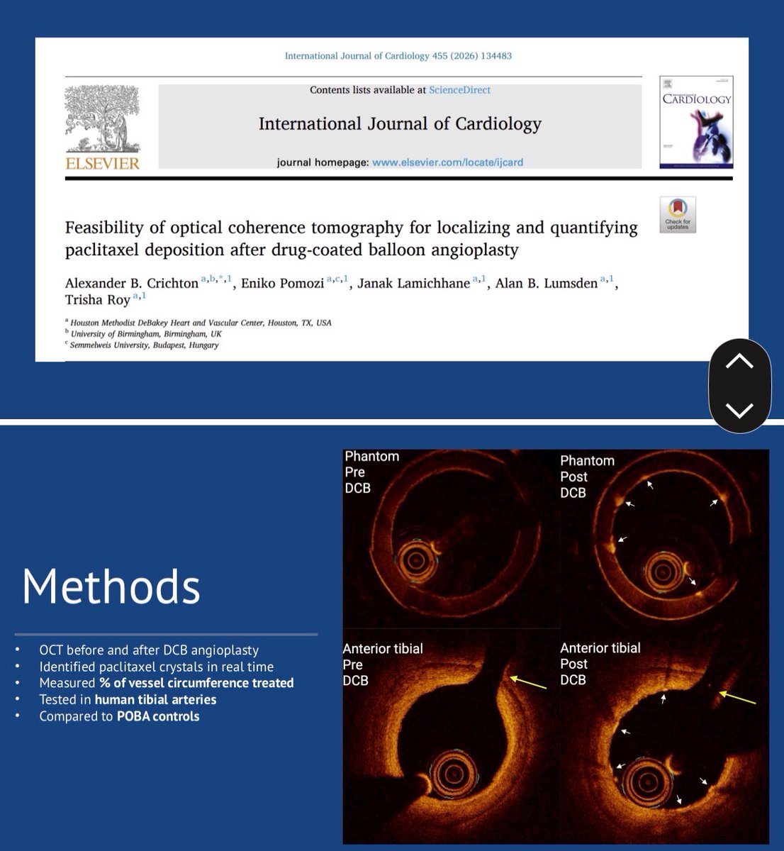
Thank you @JVascSurg for highlighting our paper this evening for the JVS journal club! Great discussion and looking forward to more collaborations in the future! @mhumphriesmd @VikKashyapMD @AeroMD @VascularABC @AlanLumsdenMD @DeBakeyCVedu


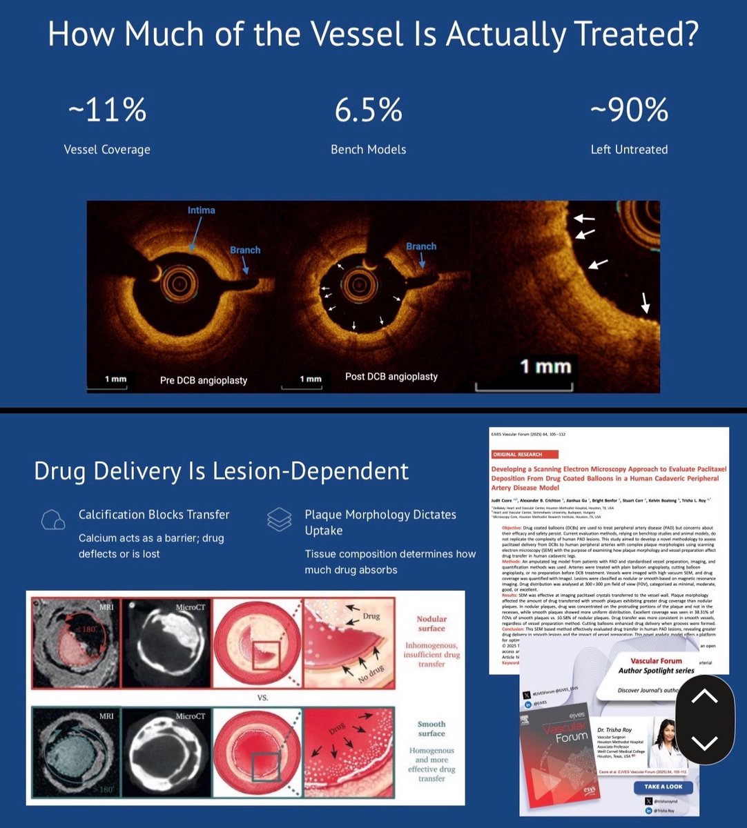
🌟 AUTHOR SPOTLIGHT: Dr. Trisha Roy 🌟 Visualizing the invisible: How drugs interact with complex plaque. In a recent #EJVESForum study, @TrishaRoyMD and her team at Houston Methodist and Weill Cornell Medical College developed a groundbreaking Scanning Electron Microscopy (SEM) approach to evaluate how paclitaxel is deposited from Drug-Coated Balloons (DCBs). 🔍 MEET THE AUTHOR: Affiliations: Houston Methodist Hospital & Weill Cornell Medical College, Houston, TX, USA 🇺🇸. Expertise: A pioneer in using advanced imaging and human cadaveric models to study peripheral artery disease (PAD) and endovascular outcomes. The Vision: Moving beyond animal models to understand how vessel preparation—like cutting balloons—changes drug transfer in real human calcified lesions. 💡 WHY THIS RESEARCH MATTERS: Current DCB data often comes from healthy animal vessels, which don't mimic the "rock-hard" calcium surgeons face daily. Dr. Roy’s work uses a human cadaveric PAD model to show that vessel preparation significantly impacts how much drug actually stays on the vessel wall. 🚀 THE CLINICAL IMPACT: By using SEM to map drug distribution at a microscopic level, we can finally see why some lesions respond to DCBs while others don't. It’s about optimizing every millimeter of the artery for the best long-term patency. Read the full study and join the discussion 👇 ejvesvascularforum.com/article/S2666-… #EJVESforum #VascTwitter #VascularSurgery #PAD #DCB #ScanningElectronMicroscopy #MedEd #ESVScommunity #VasculaRes #SurgicalInnovation #MeetTheAuthor @vascularis @bazinger_z @FLareyre @MariaKatsarou14 @NicLeoneMD @ESVSmembership @EVST_ESVS @WeillCornell @HMethodistCV @HMethodistMD @MethodistHosp

🌟 AUTHOR SPOTLIGHT: Dr. Trisha Roy 🌟 Visualizing the invisible: How drugs interact with complex plaque. In a recent #EJVESForum study, @TrishaRoyMD and her team at Houston Methodist and Weill Cornell Medical College developed a groundbreaking Scanning Electron Microscopy (SEM) approach to evaluate how paclitaxel is deposited from Drug-Coated Balloons (DCBs). 🔍 MEET THE AUTHOR: Affiliations: Houston Methodist Hospital & Weill Cornell Medical College, Houston, TX, USA 🇺🇸. Expertise: A pioneer in using advanced imaging and human cadaveric models to study peripheral artery disease (PAD) and endovascular outcomes. The Vision: Moving beyond animal models to understand how vessel preparation—like cutting balloons—changes drug transfer in real human calcified lesions. 💡 WHY THIS RESEARCH MATTERS: Current DCB data often comes from healthy animal vessels, which don't mimic the "rock-hard" calcium surgeons face daily. Dr. Roy’s work uses a human cadaveric PAD model to show that vessel preparation significantly impacts how much drug actually stays on the vessel wall. 🚀 THE CLINICAL IMPACT: By using SEM to map drug distribution at a microscopic level, we can finally see why some lesions respond to DCBs while others don't. It’s about optimizing every millimeter of the artery for the best long-term patency. Read the full study and join the discussion 👇 ejvesvascularforum.com/article/S2666-… #EJVESforum #VascTwitter #VascularSurgery #PAD #DCB #ScanningElectronMicroscopy #MedEd #ESVScommunity #VasculaRes #SurgicalInnovation #MeetTheAuthor @vascularis @bazinger_z @FLareyre @MariaKatsarou14 @NicLeoneMD @ESVSmembership @EVST_ESVS @WeillCornell @HMethodistCV @HMethodistMD @MethodistHosp


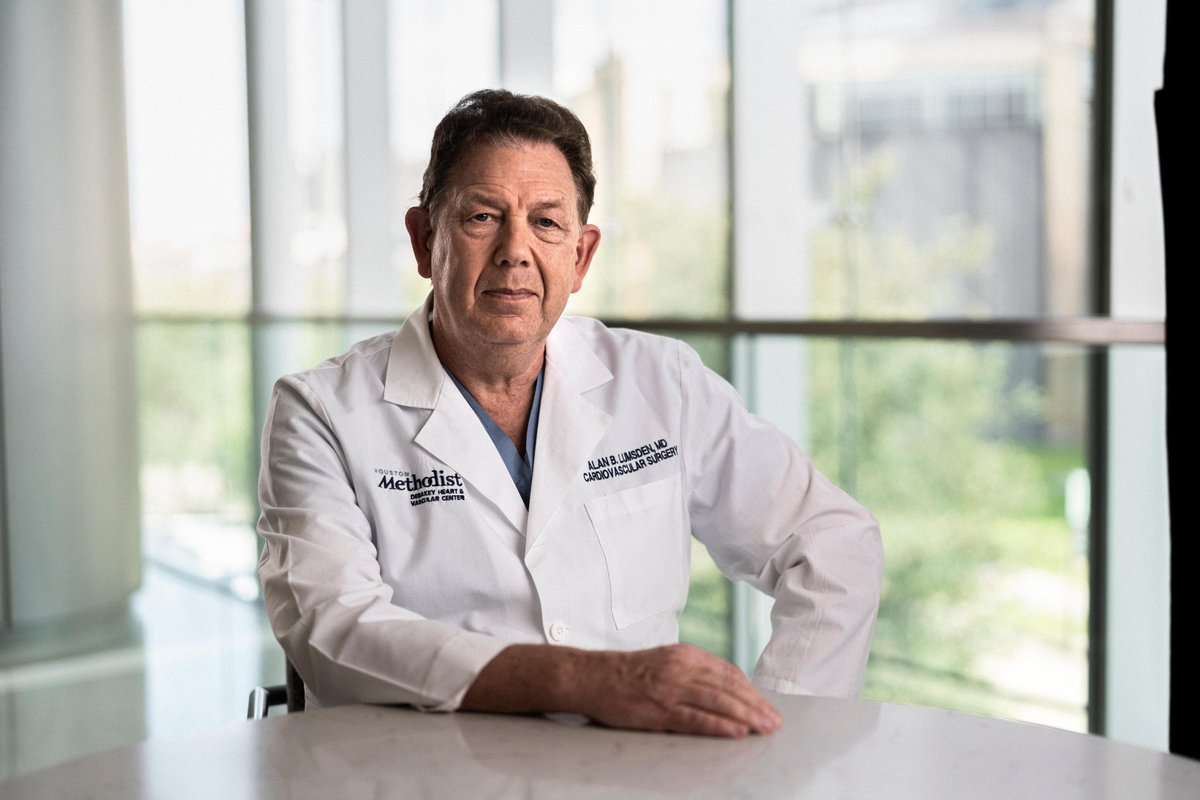
Today during Grand Rounds, Dr. Alan Lumsden shared an update on The Status of the DeBakey Heart & Vascular Center! Watch it here: bit.ly/3NnZQE3 #GrandRounds #HoustonMethodist #DeBakeyCVEducation