Ubaldo La Brocca

1.6K posts

Ubaldo La Brocca banner
Ubaldo La Brocca

Ubaldo La Brocca

@baldo_lb

🇪🇺🇮🇹 MD👨‍⚕️ Pre-Hospital Emergency 🚑 Anesthesia and Intensive Care ⚡️

Torino, Piemonte Katılım Ağustos 2011
912 Takip Edilen90 Takipçiler
Ubaldo La Brocca retweetledi
CardiovascularCorner
CardiovascularCorner@TrackYourHeart·
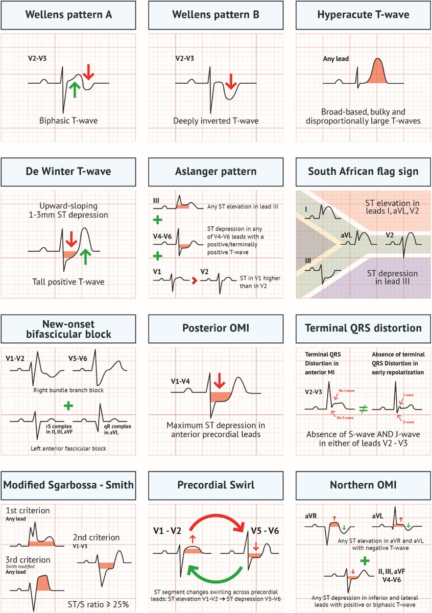
Acute MI is not always STEMI. A major 2025 review highlights why relying only on classic STEMI criteria can miss dangerous coronary occlusions. A thread on OMI (Occlusion Myocardial Infarction): 🧵
CardiovascularCorner tweet media
English
2
185
533
19.4K
Ubaldo La Brocca retweetledi
Reads with Ravi
Reads with Ravi@readswithravi·
I’m in love with this sentence: “The art of not being ready and doing it anyway will take you so far.”
English
65
2.3K
13.3K
163.8K
Ubaldo La Brocca retweetledi
Card. Gianfranco Ravasi
La sapienza non è riducibile solo a bianco e nero, non è relegabile in uno stampo prefissato né sottoponibile a un giudizio esclusivo. Per questo, senza cadere nel relativismo, si deve essere comprensivi e duttili, rigorosi ma non rigidi, consapevoli che la realtà è complessa.
Italiano
10
29
176
2.6K
Ubaldo La Brocca retweetledi
Dr. Chacón-Lozsán F .'.
Dr. Chacón-Lozsán F .'.@franciscojlk·
🧠⚡ EEG may be one of the most underused monitoring tools in modern intensive care. This new 2026 perspective in Critical Care argues something provocative: EEG should evolve from a specialist diagnostic test into a true bedside monitoring system for intensivists. The paper highlights a reality we face daily in ICU: Many critically ill patients develop: • delirium • non convulsive seizures • metabolic encephalopathy • hypoxic ischemic injury • sedation related cortical suppression Yet much of this cerebral dysfunction remains clinically invisible. Especially in: ⚠️ sedated ⚠️ mechanically ventilated ⚠️ unconscious patients Clinical examination alone is often insufficient. One of the strongest points of the article: The authors argue EEG should follow the same trajectory as: 📌 bedside echocardiography 📌 lung ultrasound Not every intensivist needs to become a neurophysiologist. But intensivists should learn to recognize: • background suppression • asymmetry • burst suppression • epileptiform activity In the same way we identify: • RV failure • tamponade • B lines • pneumothorax This is a major conceptual shift. The proposed model is particularly interesting. The authors describe a tiered EEG system: 🟢 simple bedside alarms for nurses 🟡 simplified interpretation for ICU physicians 🔵 qEEG trend analysis for trained intensivists 🔴 full raw EEG interpretation for neurophysiologists Combined with: 🤖 AI assisted pattern recognition The objective is not replacing experts. The objective is: faster recognition of dangerous brain physiology. One of the most important messages: Current ICU monitoring focuses heavily on: • blood pressure • oxygenation • cardiac output • ventilation But often ignores the organ we are ultimately trying to protect: 🧠 the brain. EEG may become the missing physiological layer of multimodal ICU monitoring. The article also raises a very practical concern: alarm fatigue. If EEG systems generate excessive false positives, ICU staff will rapidly ignore them. This is where AI may become transformative: continuous surveillance without fatigue. Particularly fascinating is the potential future of: 📡 point of care EEG 📡 emergency department EEG 📡 prehospital EEG 📡 tele neurophysiology Imagine: an ambulance transmitting simplified cerebral activity before hospital arrival. That possibility no longer sounds futuristic. My personal takeaway: Critical care spent decades refining cardiovascular monitoring. The next frontier may be: continuous functional brain monitoring. Not simply detecting seizures. But integrating cerebral physiology into real time ICU decision making. 📖 Reference Taccone, F. S., Critical Care, 30, 195. doi.org/10.1186/s13054…
Dr. Chacón-Lozsán F .'. tweet media
English
1
50
166
6.9K
Ubaldo La Brocca retweetledi
Dr. Chacón-Lozsán F .'.
Dr. Chacón-Lozsán F .'.@franciscojlk·
🫀📈 One of the most controversial debates in critical care may be shifting again. This new 2026 systematic review and meta analysis in Annals of Intensive Care challenges that narrative directly. The study analyzed: 📊 34 studies 📊 636,441 shock patients 📊 PAC, PiCCO and advanced hemodynamic monitoring guided resuscitation strategies Main finding: ✅ significant reduction in in hospital mortality with advanced hemodynamic monitoring guided management (OR 0.66) The strongest signal appeared in: ⚠️ cardiogenic shock particularly with pulmonary artery catheter guided therapy. One of the most interesting physiological observations: Patients monitored with advanced hemodynamic systems received: • more vasopressors • more inotropes • more mechanical circulatory support • more RRT Yet mortality was LOWER. That is extremely important. This suggests the benefit may not come from the device itself, but from: 🧠 earlier recognition of instability 🧠 physiology informed escalation 🧠 more precise therapeutic targeting In other words: better decision making. The paper strongly supports a concept many intensivists intuitively recognize at bedside: Not all shock is “vasoplegia plus fluids.” Different hemodynamic phenotypes require: • different vasoactive strategies • different fluid approaches • different escalation timing • different mechanical support thresholds Advanced monitoring may allow clinicians to move away from: “one size fits all resuscitation.” Another important nuance: The mortality benefit was strongest in cardiogenic shock. The evidence in septic shock remains less definitive, although trends still favored advanced monitoring. This may reflect an important reality: cardiogenic shock is fundamentally a hemodynamic disease. One particularly valuable message from this paper: The authors emphasize that modern AHDM is not simply “placing a Swan Ganz catheter.” It is: 📌 integrating dynamic physiology 📌 interpreting perfusion targets 📌 understanding ventricular interactions 📌 identifying fluid responsiveness limitations 📌 tailoring escalation Technology without physiology remains insufficient. Interesting practical point: The analysis did NOT show major increases in serious complications related to advanced monitoring devices. That matters because procedural fear has been one of the strongest arguments against invasive monitoring. My personal takeaway: Critical care may be entering a new era where: precision hemodynamics returns to the center of shock resuscitation. Not because catheters are fashionable again because modern shock management increasingly requires individualized physiology rather than protocolized averages. 📖 Reference Nagy, L., Tóth, P. R., Turan, C., et al. (2026). Annals of Intensive Care, 16, 100071. doi.org/10.1016/j.aico…
Dr. Chacón-Lozsán F .'. tweet media
English
2
57
182
8.6K
Ubaldo La Brocca retweetledi
Card. Gianfranco Ravasi
Card. Gianfranco Ravasi@CardRavasi·
Confucio ammoniva: «Non preoccuparti del fatto che la gente non ti conosce. Preoccupati piuttosto del fatto che forse non meriti di essere conosciuto. Il vero uomo soffre se è incompetente, non se è misconosciuto». Preoccupiamoci meno, allora, di fama, successo e consenso.
Italiano
8
37
296
4.2K
Ubaldo La Brocca retweetledi
Nick Mark MD
Nick Mark MD@nickmmark·
One of my favorite aviation phrases: “Superior pilots use their superior judgement to avoid having to demonstrate their superior skills.” It’s true for any proceduralist btw.
English
6
85
463
23.5K
Ubaldo La Brocca retweetledi
The Shift Journal
The Shift Journal@TheShiftJournal·
— Marcus Aurelius
The Shift Journal tweet media
CY
7
374
1.9K
48.9K
Ubaldo La Brocca retweetledi
Dr. Chacón-Lozsán F .'.
Dr. Chacón-Lozsán F .'.@franciscojlk·
🫀🤔 Rethinking renal perfusion in critical illness We often default to MAP targets, but this review challenges that simplicity. 🔑 Key insight: Renal perfusion pressure (RPP) = MAP- venous pressure (or CVP/MSFP) → Meaning both hypotension and venous congestion can drive AKI. 📌 What stands out: • The kidney is highly autoregulated… until it isn’t • In critical illness, autoregulatory failure occurs earlier than expected • AKI can develop even with “normal” macrocirculation → microcirculation matters • Fixed MAP targets ignore inter- and intra-patient variability 💡 Clinical implications: • Think beyond MAP → consider mean perfusion pressure (MPP) • Avoid venous congestion (CVP, intra-abdominal pressure, fluid overload) • Move toward individualized perfusion targets • Use multimodal monitoring (Doppler, biomarkers, tissue oxygenation) ⚠️ Bottom line: Renal protection is not just about pressure — it’s about gradients, flow, and congestion. A shift from “one-size MAP” → precision hemodynamics is coming. 📚 Reference Panwar, R., (2025) Annals of Intensive Care, 15, 115. doi.org/10.1186/s13613…
Dr. Chacón-Lozsán F .'. tweet media
English
2
40
115
5.7K
Ubaldo La Brocca retweetledi
Dr. Chacón-Lozsán F .'.
Dr. Chacón-Lozsán F .'.@franciscojlk·
🫀 Hemodynamics is not blood pressure... actually, It never was. ⚠️ The biggest mistake in perioperative & critical care: 👉 Treating numbers instead of physiology 📊 What we were taught ✔️ BP ✔️ HR ✔️ SpO₂ 🔥 What actually matters 👉 Flow + oxygen delivery + tissue perfusion 🧠 Core concept 👉 Blood pressure ≠ perfusion You can have: ▪️ Normal BP → low cardiac output ▪️ High BP → poor microcirculation ▪️ Stable vitals → ongoing hypoxia 💡 Why? Because: 👉 BP = CO × SVR Same pressure → completely different physiology 🧬 The real pillars of hemodynamics ✔️ Cardiac output ✔️ Stroke volume ✔️ Preload / afterload / contractility ✔️ Oxygen delivery (DO₂) ⚠️ Critical insight 👉 Oxygen delivery = CO × arterial O₂ content Not: ❌ BP ❌ SpO₂ alone 🔥 This is where advanced monitoring changes everything 👉 From static → dynamic 👉 From guess → prediction 🧠 Dynamic parameters outperform static ones ✔️ SVV ✔️ PPV ✔️ PVI 👉 Predict fluid responsiveness 👉 Avoid fluid overload 💥 Reality check Only ~50% of unstable patients respond to fluids 👉 The rest get harm 🫀 Next level thinking 👉 Ventriculo-arterial coupling 👉 Cardiac power output 👉 Tissue perfusion markers 🚨 Final message Stop asking: ❌ “What is the blood pressure?” Start asking: 👉 “Is the patient perfusing?” 🧠 Because in critical care: 👉 Flow saves organs Pressure just looks good on the monitor 📚 Demir et al., Aydın et al. Turkish Journal of Anaesthesiology & Reanimation, 2025 DOI: 10.4274/TJAR.2025.251926 DOI: 10.4274/TJAR.2025.251925
Dr. Chacón-Lozsán F .'. tweet mediaDr. Chacón-Lozsán F .'. tweet media
English
4
44
141
7K
Ubaldo La Brocca retweetledi
Benigno Valderrábano Salas
🫀 Did you know where the recommendation to place the pulsed wave Doppler sample volume 0.5–1 cm from the aortic valve to measure LVOT VTI comes from? The answer is more interesting than it seems. It doesn’t come from a single study or an experiment designed for that purpose. It comes from a historical chain spanning nearly 40 years: 🔬 1982–1984 — The physical foundation Pasipoularides and Murgo demonstrated using invasive catheters and mathematical models that in aortic stenosis there is a real zone of flow acceleration in the LVOT, just proximal to the valve, without any second anatomic obstruction. Pure hemodynamics — no Doppler yet. 👉 Bird et al. Circulation 1982 → doi.org/10.1161/01.CIR… 👉 Pasipoularides et al. Am J Physiol 1984 → doi.org/10.1152/ajphea… 📐 1984 — The apical 5-chamber view Lewis, Kuo and Quinones were the first to validate cardiac output measurement using pulsed wave Doppler from the cardiac apex. They described placing the sample volume “immediately proximal to the aortic valve leaflets” — but without specifying any distance in centimeters. 👉 Lewis et al. Circulation 1984 → doi.org/10.1161/01.CIR… 📏 1985 — The first numerical distance Skjaerpe, Hegrenaes and Hatle (the Norwegian group) were the first to quantify this in Doppler: they empirically observed that flow acceleration began 0.5 to 1.5 cm proximal to the valve, and placed the sample volume just proximal to that zone. They directly cited Pasipoularides as supporting evidence. This was the first time a numerical distance appeared in the technique. 👉 Skjaerpe et al. Circulation 1985 → doi.org/10.1161/01.CIR… 📊 1986–1988 — Practical consolidation Otto et al. used ~1.0 cm. Oh, Tajik and the Mayo Clinic group explicitly established the range of 0.5 to 1.0 cm in 100 patients, justifying it as necessary to avoid the subvalvular acceleration zone. This is the figure we all recognize today. 👉 Otto et al. JACC 1986 → doi.org/10.1016/S0735-… 👉 Zoghbi et al. Circulation 1986 → doi.org/10.1161/01.CIR… 👉 Oh et al. JACC 1988 → doi.org/10.1016/0735-1… 📋 2002 — It becomes “official” Quinones, Otto, Zoghbi and colleagues codified it in the ASE guidelines as “~5 mm proximal to the aortic valve”… but without citing any specific study to support it. It had already become expert consensus. 👉 Quiñones et al. JASE 2002 → doi.org/10.1067/mje.20… ⚔️ 2017 — The debate reopens Baumgartner et al. (EACVI/ASE) maintained the 0.5–1 cm recommendation. However, Hahn and Pibarot responded with a critical letter pointing out that the original articles from the 1980s measured at the aortic annulus, not 0.5–1 cm below it, and that moving away from the annulus introduces errors due to the elliptical and irregular shape of the subannular LVOT. 👉 Baumgartner et al. Eur Heart J Cardiovasc Imaging 2017 → doi.org/10.1093/ehjci/… 👉 Hahn & Pibarot. JASE 2017 → doi.org/10.1016/j.echo… 💡 Bottom line: The 0.5–1 cm figure was never experimentally validated as the optimal distance. It emerged from empirical observations in the 1980s aimed at avoiding a flow acceleration zone that had been demonstrated with invasive catheters. It was adopted through accumulated clinical practice and later elevated to a formal recommendation by consensus. The debate over whether to measure at the annulus or 0.5–1 cm below it remains open to this day. One of those recommendations we all follow but few know where it actually came from 🙂 Dr Benigno Valderrábano Salas @MDBeni @JaeKOh2 @ottoecho @WilliamZoghbi @ASE360 @EACVIPresident @NephroP @iamritu @PPibarot @hahn_rt @MAecocardio @SISIACOficial @SONECOM_AC @VazyurVasquez @Cardiotweets83 @HEARTof_echo @echobasics
Benigno Valderrábano Salas tweet media
English
17
133
411
53.9K
Ubaldo La Brocca retweetledi
Brian Locke, MD MSCI
Brian Locke, MD MSCI@doc_BLocke·
“Stewart Light” approach to acid-base and visualization Classical ABG interp: use pH, PaCO₂, HCO₃⁻ [or SBE, base excess], and the Boston compensation rules to decide whether the respiratory response fits. It works well for single disturbance, but incomplete when several coexisting processes. For eg - if SBE = 0, there could be no metabolic disturbance, or two that are equal but opposite. Stewart/Physiochemical approach = three indep vars: PCO2, strong ion difference (SID), and total weak acid concentration ([ATOT].. mainly alb and phos) In “A pragmatic approach to complex acid base disturbances of critical illness: the ‘Stewart light’” the authors propose a hybrid approach: - keep Boston rules for respiratory/compensation - add pH-adjusted partition of metabolic SBE into chloride/SID effect (from Na - Cl), albumin effect, and unmeasured-ion reminder Why? To tease out situations where multiple metabolic processes pull in opposite directions, for e.g. when a Strong Ion Difference and unmeasured-ion (or lactate) coexist. Most useful for DKA, intoxication, sepsis, vomiting, NS administration, diuresis, or [my interest] chronic hypercapnia… Boston Rules can tell if you if the net secondary response(s) are as expected, but doesn’t tell what type of metabolic processes are present. 5 steps: 1. clinical context, pH, severity, and Boston rules (e.g. winters formula for met acidosis) 2. Quantify strong-ion effect SBE_SID = Na⁺ − Cl⁻ − 35; if pH is outside 7.30–7.50, adjust that reference 35 by 1.5 mmol/L for each 0.1 pH unit away from 7.40—upward in acidemia, downward in alkalemia. Negative values = strong-ion acidosis, positive values = stron-ion alkalosis 3. Quantify weak-acid effect from albumin SBE_Alb = 0.3 × (40 − albumin in g/L), so hypoalbuminemia is treated as an alkalinizing force. 4. Assign remainder: unmeasured ions: SBE_UI = SBE − SBE_SID − SBE_Alb; if lactate is available, subtract its contribution to see whether additional unmeasured anions remain Then, 5.: synthesize all together. (and clinically determine if a positive SBE_SID is due to chloride depletion primary process or expected renal repsonse to chronic hypercapnia). They recommend visualizing it with a gamblegram and practicing… and this all seems to hard. So I vibe coded a visualization, available here: Takes your blood gas and chemistries result -> gives “Stewart Light” interpretation and some visualizations/explanations.
Brian Locke, MD MSCI tweet mediaBrian Locke, MD MSCI tweet mediaBrian Locke, MD MSCI tweet media
English
2
85
285
29.6K
Ubaldo La Brocca retweetledi
Dr. Chacón-Lozsán F .'.
Dr. Chacón-Lozsán F .'.@franciscojlk·
🫁 Why I always ask for paired blood gases! CO2 and hemodynamics 🧪 For years, we have relied on: ▪️ Lactate ▪️ ScvO₂ / SvO₂ ▪️ Clinical perfusion But all of them share a critical limitation: 👉 They do not reliably detect ongoing tissue hypoperfusion ⚠️ The problem You can have: ✔️ Normal ScvO₂ ✔️ Decreasing lactate ✔️ “Stable” hemodynamics …and still have microcirculatory failure 👉 This is where CO₂ enters the game 🧠 The physiology in short CO₂ behaves differently from oxygen: ➡️ ~20x more diffusible than O₂ ➡️ Accumulates when flow is insufficient ➡️ Reflects flow adequacy, not just oxygenation 👉 Pv-aCO₂ ≈ inverse of cardiac output 🔥 What the CO₂ gap really tells you 🟢 Pv–aCO₂ < 6 mmHg → Likely adequate flow 🔴 Pv–aCO₂ ≥ 6 mmHg → Suggests low flow / impaired perfusion BUT: ❗ It is NOT a marker of hypoxia alone ❗ It is a marker of flow–metabolism mismatch ⚡ The real upgrade: the CO₂/O₂ ratio 👉 Pv-aCO₂ / Ca-vO₂ This is the missing piece. ✔️ Approximates respiratory quotient ✔️ Detects anaerobic metabolism ✔️ Reacts faster than lactate 📈 >1 = ongoing anaerobic metabolism 🚨 Clinical implications 🩸 Septic shock High CO₂ gap despite ScvO₂ >70% → hidden hypoperfusion Persistent Pv–aCO₂ ≥6 mmHg → ↑ mortality 🫀 Fluid responsiveness ↓ Pv–aCO₂ after fluids → likely responder 🫁 Weaning failure ↑ CO₂ gap during SBT → inadequate DO₂ vs VO₂ 🏥 Post-op patients Elevated CO₂ gap predicts complications better than lactate ❌ Common mistakes ❌ Using lactate alone ❌ Ignoring normal ScvO₂ “false reassurance” ❌ Interpreting CO₂ gap without context (pH, Hb, ventilation) ❌ Treating numbers instead of physiology 🚀 Modern hemodynamic approach We should integrate: 1. Macrocirculation → MAP, CO 2. Oxygen markers → ScvO₂ 3. Metabolic markers → Lactate 4. Flow markers → Pv–aCO₂ 5. Anaerobic markers → Pv–aCO₂ / Ca–vO₂ 👉 Not one variable 👉 A physiology-driven bundle 🎯 Take-home CO₂ is not a waste product. 👉 It is a real-time marker of perfusion adequacy 👉 It detects what oxygen variables miss 👉 It bridges macro and microcirculation 📚 Mallat J et al. (2025) Annals of Intensive Care DOI: 10.1186/s13613-025-01569-2
Dr. Chacón-Lozsán F .'. tweet media
English
6
112
353
30.4K
Ubaldo La Brocca retweetledi
Card. Gianfranco Ravasi
Card. Gianfranco Ravasi@CardRavasi·
Ai giovani: non temete il flusso degli anni, perché esso porta con sé esperienza, sapienza, consiglio e, quindi, migliora la persona. Ogni età è bella purché l'anima (e non tanto il corpo) abbia poche rughe.
Italiano
10
45
345
5.2K
Ubaldo La Brocca retweetledi
Kelly Haughton, MD
Kelly Haughton, MD@kidneydoctorpa·
AKI guidelines hadn’t been updated since 2012. The KDIGO 2026 AKI/AKD Public Review Draft just dropped and it changes how we define, diagnose, and follow up after acute kidney injury. Here’s what every nephrologist, intensivist, and internist needs to know 🧵 ⚠️ Public review draft only · Not yet final guidelines
English
14
407
1.3K
294.1K
Ubaldo La Brocca retweetledi
Fabio Ferrero
Fabio Ferrero@BiettoFerrero·
Thanks @Tesla and @Tesla_AI for allowing me to test drive FSD in Turin this morning. It was during Auto Moto Turin Show, and to my eyes all other thousands of ICE car there looked like centuries in the past, like I was in a mausoleum. Can’t wait for EU approval! 🇪🇺
English
0
1
1
37
Ubaldo La Brocca retweetledi
Reads with Ravi
Reads with Ravi@readswithravi·
This paragraph by Richard Feynman hits so hard: “Fall in love with some activity, and do it! Nobody ever figures out what life is all about, and it doesn’t matter. Explore the world. Nearly everything is really interesting if you go into it deeply enough. Work as hard and as much as you want to on the things you like to do the best. Don’t think about what you want to be, but what you want to do. Keep up some kind of a minimum with other things so that society doesn’t stop you from doing anything at all.”
English
60
1.1K
6.3K
216.9K
Ubaldo La Brocca retweetledi
Your Best Version
Your Best Version@YourPrimePath·
A man’s greatest weapon is consistency. It outlasts talent, talk, and luck.
English
96
1.5K
8.5K
121.6K
Ubaldo La Brocca retweetledi
Chris Bishop
Chris Bishop@Pipes_n_pumps·
The global oxygen delivery (DO2) formula - critical to understanding and approaching “big sick” patients This was something I learnt very early on in my clinical perfusion career and one of the first things I teach trainees and students - whether the pump is the native heart or extracorporeal pump, the principle is the same
Chris Bishop tweet media
Haney Mallemat@CriticalCareNow

Cardiogenic shock and the DO2 formula The oxygen delivery formula is the only math that matters in a crash! DO2 depends on your Hgb, O2 Sat, and Cardiac Output. Don't be fooled by 100% SpO2—if the pump isn't circulating that "cargo," the organs are still starving.

English
3
46
168
17.3K