Pedro Rojas

2.6K posts

Pedro Rojas banner
Pedro Rojas

Pedro Rojas

@pedrorojma

Médico cirujano ortopedista

Puebla, México Katılım Ocak 2010
1.1K Takip Edilen232 Takipçiler
Pedro Rojas retweetledi
Jeremy Burnham, MD
Jeremy Burnham, MD@JeremyBurnhamMD·
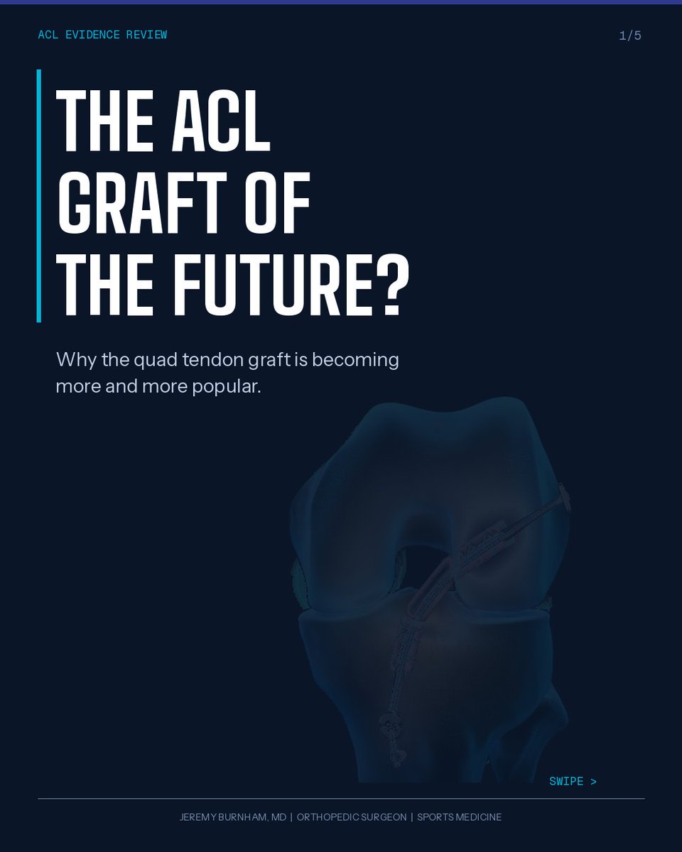
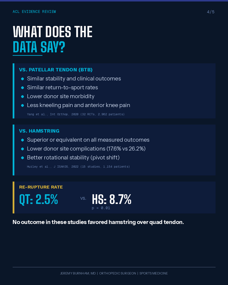
Quad tendon: 2x the cross-sectional area of patellar tendon, 38% greater load to failure, 68% greater stiffness, and less harvest-site pain (Shani et al., 2016). Re-rupture rate: QT 2.5% vs. hamstring 8.7%, p=0.01 (Hurley et al., 2022). A meta-analysis of 2,962 patients across 32 RCTs found QT superior to both BTB and hamstring on key outcome measures (Yang et al., 2020). BTB is still a great graft, but the data on QT keeps getting stronger.
Jeremy Burnham, MD tweet mediaJeremy Burnham, MD tweet mediaJeremy Burnham, MD tweet mediaJeremy Burnham, MD tweet media
English
9
5
41
6.3K
Pedro Rojas retweetledi
Mo Imam
Mo Imam@MoAImam·
The rotator cuff muscles seen from above
Mo Imam tweet media
English
1
11
51
1.4K
Pedro Rojas retweetledi
Dr. Mateo Avila
Dr. Mateo Avila@drteoa·
¿Sigues pidiendo TP, TTPa y plaquetas a TODOS los pacientes antes de operar “por rutina”? Un consenso Delphi 2025 con anestesiólogos cuestiona esta práctica y propone algo mucho más lógico. Te cuento los puntos clave 👇
Dr. Mateo Avila tweet media
Español
12
276
1.5K
79.2K
Pedro Rojas retweetledi
Antelm Pujol, MD PhD
Antelm Pujol, MD PhD@AntelmPujol·
🤧🤧 ¿EJERCICIO FÍSICO PARA LA PREVENCIÓN DEL CATARRO COMÚN/ RESFRIADO? 🏃‍♀️ La realización de ejercicio físico 5 veces por semana se asocia: 📉43-46% MENOS probabilidad de resfriarte. 📉32-41% REDUCCIÓN de la severidad. 📉34-41% REDUCCIÓN de la intensidad de los síntomas.
Antelm Pujol, MD PhD tweet media
Español
3
174
597
22.1K
Pedro Rojas retweetledi
Dr. Shubham Arora
Dr. Shubham Arora@Dr_S_Arora·
I’m elated to announce the launch of our first website. This platform brings together everything I care deeply about: radiology education, medical illustration, and creating visual tools that make learning simpler radiologyillustration.com
Dr. Shubham Arora tweet media
English
13
112
735
38K
Pedro Rojas retweetledi
Jeremy Frisch
Jeremy Frisch@JeremyFrisch·
The Youth athletic development starter kit: Pool noodles Flags Tennis balls Traffic cones 2x4s Pvc Mini-hurdles Jump ropes You don't need a multi-million dollar training facility to deliver world class athletic development. #LTAD
English
0
37
258
15.8K
Pedro Rojas retweetledi
Gerry DeFilippo
Gerry DeFilippo@Challenger_ST·
8 plyometrics to enhance your athleticism! 1. Quadruple broad jump 2. SL hurdle to lateral jump 3. Depth jump to broad jump 4. Repeat maximal pogo jump 5. “Double,” box jump 6. SL drop jump 7. Lateral jump to 1/4 turn box jump 8. Maximal skater jump
English
6
95
961
68.9K
Pedro Rojas retweetledi
sergio serrano belmar
sergio serrano belmar@sserranobmsk·
Sternoclavicular joint instability: diagnosis and treatment using ultrasound A 24-year-old female rugby player, after making a tackle, began experiencing pain in the superomedial region of the right hemithorax. This pain did not prevent her from continuing the game, but forced her to shift her tackle to the left side. On physical examination, she presented with tenderness to palpation of the right sternoclavicular joint, with positive adduction tests against resistance and active compression. The video initially shows the left sternoclavicular joint, revealing a preserved joint space associated with a normal anterior sternoclavicular ligament, with usual joint mobility during dynamic compression of the distal end of the clavicle. Subsequently, the right sternoclavicular joint is visualized with significant synovial thickening within it, associated with a complete tear of the anterior sternoclavicular ligament at the clavicular end, and anterior subluxation of the clavicle during dynamic assessment. Non-surgical treatment is decided upon by resting the joint with a sling, followed by intra-articular infiltration of 1 cc of triamcinolone and 0.5 cc of 2% lidocaine for pain control, and subsequently starting rehabilitation.
English
0
8
39
3.2K
Pedro Rojas retweetledi
Rad_AML ☢️
Rad_AML ☢️@Rad_AML·
ULTRASOUND CHRONICLES (Episode 3) Topic: Basic Physics of Ultrasound (Part II) Every image begins as sound; bouncing, bending, and fading through the body. Understanding how these waves behave turns echoes into stories. 1. ACOUSTIC IMPEDANCE AND IMAGE FORMATION Every tissue has a unique sound personality (its acoustic impedance, Z). This is the resistance it offers to sound (density × velocity). When sound meets tissues with different impedances, part reflects to form echoes while the rest continues forward. A greater difference = stronger reflection (brighter echo). A smaller difference = weaker reflection (darker echo). Example: Liver-diaphragm (bright) vs. Liver-kidney (smooth). Clinical Correlate: Image brightness reflects impedance difference. 2. ULTRASOUND RESOLUTION Resolution defines how well two close points are seen as separate. 1. Axial: Along beam path; improved by higher frequency and shorter pulses. 2. Lateral: Side by side; best at the focal zone. 3. Elevational: Slice thickness depends on beam width and probe design. ➡️Good resolution = crisp detail. ➡️Poor resolution = blurred or merged structures. Clinical Correlate: Two small cysts appearing as one? It’s likely a resolution issue, not pathology. 3. IMAGE DISPLAY IN ULTRASOUND (ECHOGENICITY) Ultrasound images appear in shades of gray, with each shade telling how much sound a tissue reflects. A) Anechoic: No echoes (black); fluid-filled (e.g, urinary bladder, gallbladder, or simple cyst). B) Hypoechoic: Few echoes (dark gray); soft tissues (e.g, renal cortex or lymph nodes). C) Isoechoic: Same brightness as surroundings (e.g, liver lesions blending with liver parenchyma). D) Hyperechoic: Bright echoes; dense or fibrous tissues (e.g, diaphragm, fatty liver, or gallstones). E) Complex: Mixed echoes; solid and cystic (e.g, ovarian complex cysts or abscesses). Clinical Correlate: Before chasing pathology, master gray-scale language. It’s the foundation of interpretation. 4. ULTRASOUND CONTROLS Knowing your control panel is power. These settings shape how sound is displayed. i) Gain: Brightness. ii) Depth: How deep the beam travels. iii) Focus: Sharpness at your target zone. iv) Dynamic Range: Adjusts contrast (high = smooth; low = sharp). v) Frequency: High for detail, low for depth. vi) Zoom: Magnifies the area of interest. Clinical Correlate: Balance is key. Overusing gain or TGC can mask subtle findings. 5. TIME GAIN COMPENSATION (TGC) As sound travels deeper, echoes weaken. TGC amplifies deeper echoes, keeping brightness uniform from near to far field. ➡️Flat TGC = balanced brightness. ➡️Over-steep = deep tissues too bright. Clinical Correlate: Always review TGC after changing depth; overcorrection may hide small lesions. 6. PATTERN RECOGNITION IN ULTRASOUND Beyond physics lies perception; the art of recognizing patterns. Look for echotexture, borders, shape, and posterior features like enhancement or shadowing. Examples: ▪︎Posterior enhancement = cyst. ▪︎Shadowing = gallstone. ▪︎Heterogeneous texture = inflammation or tumour. Clinical Correlate: Pattern recognition is where science becomes sight, where echoes turn into diagnosis. CONCLUSION From sound to signal, ultrasound is the poetry of physics in motion. Master these basics, and every image you capture will speak with clarity, confidence, and purpose. Bye for now 👋 @iamayims @_SusuAbdul @Rad_Munagi
Rad_AML ☢️ tweet mediaRad_AML ☢️ tweet mediaRad_AML ☢️ tweet mediaRad_AML ☢️ tweet media
Rad_AML ☢️@Rad_AML

🎬 TRAILER... ULTRASOUND CHRONICLES (Episode 3) Title: Basic Physics of Ultrasound (Part II) In the last episode, we uncovered how sound travels and forms images. Tonight, we go deeper into what shapes those images: brightness, sharpness, and control. Drops tonight || 9 pm. Stay tuned and turn on your notifications. 🔔 #UltrasoundChronicles #Rad_AML #MedicalImaging #UltrasoundPhysics

English
9
63
216
15.6K
Pedro Rojas retweetledi
Rad_AML ☢️
Rad_AML ☢️@Rad_AML·
ULTRASOUND CHRONICLES (Episode 2) Topic: Basic Physics of Ultrasound (Part I) 1. INTRODUCTION Every great image begins with one thing... sound. Understanding the physics behind ultrasound helps you control your image, troubleshoot artefacts, and appreciate the beauty of how invisible waves create visible detail. 2. CONCEPTS OF SOUND Sound is a mechanical vibration that travels as longitudinal waves through a medium (alternating areas of compression and rarefaction). It needs a medium (like tissue) to move and travel poorly through air or bone. Key terms: ▪︎ Frequency (Hz): Number of waves per second. ▪︎ Wavelength (λ): Distance between compressions. ▪︎ Velocity (v): Speed in tissue (~1540 m/s in soft tissue). ▪︎ Amplitude: It is the wave strength. Determines brightness on the screen. Medical ultrasound operates between 1-15 MHz (this is well above the human hearing range). 3. GENERATION OF SOUND Inside every transducer, electrical energy is converted into mechanical vibrations. These vibrations create pulses of sound that travel into tissue and return as echoes (this the basis of image formation). 4. THE ULTRASOUND BEAM The ultrasound beam is made up of sound pulses emitted from the transducer. It's just like a flashlight that’s narrow and bright near the source but widens with distance. It has three main zones: ▪︎ Near Field (Fresnel): Converging beam with the best resolution. Ideal for superficial scans like the thyroid or carotid. ▪︎ Focal Zone: Point of maximum clarity. Place it at your area of interest (e.g., gallbladder wall or vessel lumen) for sharp images. ▪︎ Far Field (Fraunhofer): Diverging beam with reduced sharpness. Deep organs like kidneys appear less detailed here. NB: Always position your focal zone at or just below the structure you’re examining for the clearest image. 5. TRANSDUCERS (PROBE) A transducer sends and receives sound waves (like a loudspeaker and microphone in one). Types & Frequency Ranges: ▪︎ Linear (7.5-15 MHz): High frequency for superficial scans, e.g., thyroid, breast, vascular, MSK. ▪︎ Curvilinear (2-5 MHz): Deeper penetration for abdominal, pelvic, and obstetric imaging. ▪︎ Phased Array (1.5-4 MHz): Small footprint for cardiac and intercostal scans. NB: ➡️ High frequency = detail. ➡️ Low frequency = depth. Pick your probe based on the target, not the habit. 6. PIEZOELECTRIC EFFECT This is the heart of ultrasound technology. Certain crystals (e.g., lead zirconate titanate) vibrate when electrically stimulated, producing sound waves. When echoes return, they generate voltage, converting sound back into an image. 7. INTERACTION OF ULTRASOUND WITH MATTER When sound meets tissue, the following occur: ▪︎ Reflection: Echoes form the image (e.g., diaphragm-liver interface or gallbladder wall producing strong echoes). ▪︎ Refraction: Beam bends at tissue boundaries, possibly causing displacement artefacts (e.g., at the rectus muscle-fat interface or across the bladder wall). ▪︎ Scattering: From irregular tissues adds texture is to organ parenchyma (e.g., heterogeneous appearance of the liver or renal cortex). ▪︎ Absorption: Energy turns to heat. This is the main cause of attenuation (e.g., in muscle tissue where sound energy is mostly absorbed). ▪︎ Attenuation: Beam weakens with depth, limiting penetration (e.g., difficulty visualizing deep structures in obese patients or with high-frequency probes). Each interaction shapes how bright, clear, and deep your final image appears. CONCLUSION Physics is the heartbeat of every scan. Understand it, and your images will always speak clearly. Bye for now 👋
Rad_AML ☢️ tweet mediaRad_AML ☢️ tweet mediaRad_AML ☢️ tweet mediaRad_AML ☢️ tweet media
Rad_AML ☢️@Rad_AML

Ultrasound Chronicles (UC) - Episode 2 Tonight ⏰ 9PM (WAT) 🎯 Topic: Basic Physics of Ultrasound From sound waves to image formation. We’re diving into the science that powers every scan. Stay tuned! It’s going to be illuminating 💡 @_SusuAbdul @RAD_IB_UMAR @iamayims @Rad_Munagi #UltrasoundChronicles #Rad_AML #Radiology #Sonography #MedicalImaging

English
4
58
175
20.3K
Pedro Rojas retweetledi
Gonzalo Serrano-Belmar.
Gonzalo Serrano-Belmar.@GSERRANOB_MSK·
🌀 Dynamic Anterior Instability of Discoid Lateral Meniscus Dynamic ultrasound enables us to visualize the translation of the thickened discoid meniscus during knee flexion and extension — a key finding often overlooked on static MRI. ⚡️ This anterior instability occurs when the anterior meniscocapsular attachments are lax or deficient, or torn, allowing the meniscus to glide and snap dynamically. Real-time ultrasound perfectly captures this motion, providing both diagnostic and surgical insight. 🦵✨ 🎯 Tip: Observe the anterior horn while flexing the knee — sudden displacement or “snapping” confirms anterior instability. #mskrad #mskradiology #mskultrasound #mskradiologybullets #radres
English
3
18
86
3.5K
Pedro Rojas retweetledi
Specialized Training™️
🔝"This review analyzed the available criteria to clear professional football players to RTP following an HSI, predicting lay-off time, and formulating an accurate RTP at the time of injury ⚽️" 👉 @paolop_physio @MarcoBeato1 et al, 2025 🇮🇹 📂Open Access: #abstract" target="_blank" rel="nofollow noopener">tandfonline.com/doi/full/10.10…
Specialized Training™️ tweet media
English
3
19
65
5.4K
Pedro Rojas retweetledi
Medical Education
Medical Education@MedEd_Cases·
The lateral patellar plica is a fold of synovial tissue located on the lateral aspect of the knee joint, running along the inner surface of the lateral patellar facet. Its function is generally considered minor, serving as a synovial fold that allows smooth gliding of the patella during knee movement. Clinically, it becomes significant when thickened or inflamed, potentially causing lateral knee pain, catching, or symptoms of plica syndrome, which may require conservative or surgical management. Read: wikism.org/Lateral_Patell… #Anatomy #AnatomyLearning #AnatomyEducation #AnatomyOfTheDay #SportsMedicine #Orthopedics #PhysicalTherapy #AthleticTraining #Rehab #InjuryPrevention #Physio #SportsInjury #SportsRehab #PhysioTherapy #Meded #FOAMed
Medical Education tweet mediaMedical Education tweet mediaMedical Education tweet mediaMedical Education tweet media
English
1
34
164
5.6K