suraj

62 posts

suraj banner
suraj

suraj

@surajk23

Neuroscientist Studying Circuits of Social Communication. Husband.

Washington DC Katılım Aralık 2010
608 Takip Edilen116 Takipçiler
suraj
suraj@surajk23·
@ReheSamay What have you done samay. Now this will be used to censor and curb YouTube and we will lose whatever little bit of journalism is left in the country! Thanks!!
English
0
0
0
46
suraj retweetledi
suraj retweetledi
Cell Reports
Cell Reports@CellReports·
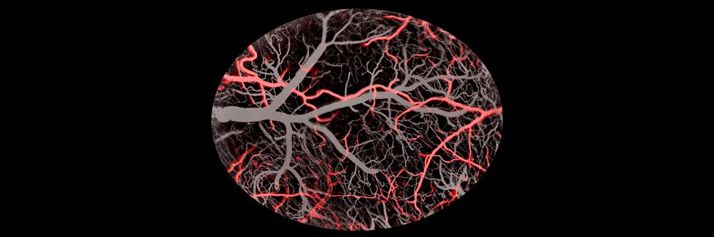
Development of pial collaterals by extension of pre-existing artery tips dlvr.it/TDg4px

English
0
4
28
13.2K
suraj retweetledi
PrakashLab
PrakashLab@PrakashLab·
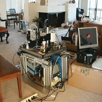
Final presentations from this years #FrugalScience2024 class are online. We cover refugee health/shistosomiasis/preparing for climate change and more.. Every year - this global community restores my passion for frugal solutions and scale. Help if you can frugalscience.org/2024-projects
English
1
8
32
6.5K
suraj retweetledi
Eric Betzig
Eric Betzig@Eric_Betzig·
#FluorescenceFriday #cellbiology #neuroscience Oligodendrocytes fluidly migrating along their thin projecting neurites in the hindbrain of a developing zebrafish embryo, as seen by adaptive optical two photon microscopy. Ultimately, these neurites form myelin sheaths that surround and insulate axons to increase the speed and efficiency of signal transmission between neurons. doi.org/10.1038/nmeth.…
English
3
34
159
17.8K
suraj retweetledi
allen institute
allen institute@AllenInstitute·
The fireworks in your mind. 🧠✨ This sparkling video shows the neurotransmitter glutamate being released into synapses, made possible by an indicator developed by @abhi_aggarwal1, @PodgorskiLab, and team. #HappyNewYear #NYE
English
11
271
1K
95.1K
suraj retweetledi
Danielle Beckman
Danielle Beckman@DaniBeckman·
Why is it so hard to investigate viral infection in the human brain? At the beginning of the #COVID19 pandemic, many studies were categorical, affirming that the virus had no neurotropism and could not infect brain cells. Many people must realize that working with postmortem human brain tissue offers many challenges for viral studies. Each human dies in different circumstances, and having the same time interval to start preparing the brain for analysis is impossible. Not only is the time variable, but it is usually very long compared to the controlled environment when working with animal models in a laboratory. In addition, several experimental models of neurotropic viral infections have shown that viruses are usually quickly cleared from the brain despite the initial fast spreading. Given that most autopsy studies inevitably have long, variable postmortem intervals before tissue processing, especially during the pandemic, infected neurons from highly connected olfactory regions may be dying early and thus evading detection. Detecting active viruses in the brain, plasma, and imaging biomarkers can help us shed light on early neurological events. Many biomarker studies have reported that cognitive decline is a common finding, and even non-severe COVID-19 is associated with early-onset cognitive decline and fluid biomarkers alteration, such as higher plasma Neurofilament concentration. There are other challenges to face regarding having good animal models for studying #SARSCoV2. The mouse ACE2 receptor does not effectively bind the viral spike protein, and Labs had to create transgenic mice models expressing human Ace2 to visualize the viral damage in the brain and other regions. I work with monkeys, which offers a substantial advantage since they can be naturally infected with SARSCoV2 and experience disease progression similar to humans. However, I am aware that most Labs don't have access to newly developed transgenic mice models or primates, and these factors contribute to slowing down and misguiding research progression in this field. For me, the damage and infection in the brain is directly visible in the microscope. We see that SARSCoV2 infects almost all cell types in the brain. We detect neurons and glia cells containing abundant Ace2 receptors and observe Spike protein (🟣) binding to them. We also observe viral double-stranded RNA (dsRNA🔴) concentrate exclusively in the cell body of neurons (neuronal protein 🟢). That means the virus can infect many different brain cell types but prefers using neurons to replicate and propagate in the brain. This affinity is likely because replicating inside the neuron and using axonal transport for propagation is the fastest mechanism for a virus to spread in the brain. Beaded, fragmented, and degenerating axons are common findings in COVID-19-infected brains; consequently, substantial degradation of myelin and neuroinflammation is also visible. Neurovirology is a recent field and is full of many challenges. Detection of single viral particles in axons or viral reproduction apparatus within neurons in brain regions remains a tremendous challenge. However, not acknowledging that SARSCoV2 infects the brain and causes substantial behavioral and cognitive alterations is denialism and irrational action to avoid the uncomfortable truth that the scientific method suggests. History has shown that this contributes to nothing other than slowing progress and discoveries, and this concept is termed the Semmelweis reflex. We need to keep fighting against it.
Danielle Beckman tweet media
English
52
328
955
145.5K
Danielle Beckman
Danielle Beckman@DaniBeckman·
A large blood vessel crosses the prefrontal cortex! Astrocytes outlining the blood vessel (🟢GFAP), filled with red blood cells (🔴RBC), and surrounded by many neurons (🟣NeuN). Happy #MicroscopyMonday! 🔬🤓#neuroscience @zeiss_micro
Danielle Beckman tweet media
English
11
56
377
27.6K
suraj retweetledi
Veera Rajagopal 
Veera Rajagopal @doctorveera·
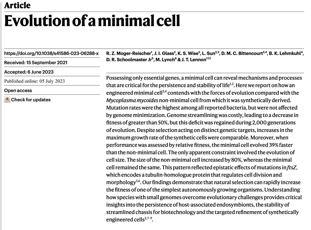
A mind-blowing paper has come out today in @Nature In 2016, JC Venter Institute scientists trimmed a bacterial genome to its barest minimum required for life to synthesize what they called a "minimal genome" (science.org/doi/10.1126/sc…). Today, a group of scientists from Indiana University reports how that minimal genome evolved over 2000 generations in comparison to the non-minimal genome. The authors found that even when you reduce a bacterial genome to its absolute minimum where every nucleotide matters, the genome undergoes mutational events generation after generation as much as the non-minimal genome. One simply cannot stop the evolution. Just over 300 days of evolution (equivalent to 40,000 years in humans) the minimal cell has gained everything it lacked in fitness on day one in comparison to the non-minimal cell. When comparing the evolved traits between the minimal and non-minimal cells, the scientists found something striking. The evolutionary process increased the cell size of non-minimal cells but not that of the minimal cell. But that is not the striking part. The scientists were able to identify the key mutation that resulted in cell size evolution. And it turned out that the mutation that helped the non-minimal cells to grow bigger is the same that helped the minimal cells to stay smaller. Growing bigger had a survival advantage for non-minimal cells and not growing bigger had a survival advantage for minimal cells. So, the mutation had a context-dependent effect. This just demonstrates that the evolutionary effects on traits have no absolute direction. All that matter is what is beneficial for the organism's survival. The conclusion of the paper is metaphorically a quote from the Jurassic Park movie: “Listen, if there’s one thing the history of evolution has taught us is that life will not be contained. Life breaks free. It expands to new territories, and it crashes through barriers painfully, maybe even dangerously, but . . . life finds a way". (scienmag.com/artificial-cel…) nature.com/articles/s4158…
Veera Rajagopal  tweet media
English
210
5.4K
20.3K
4.2M