Prof. Dr. Sanjeev Bagai@BagaiDr
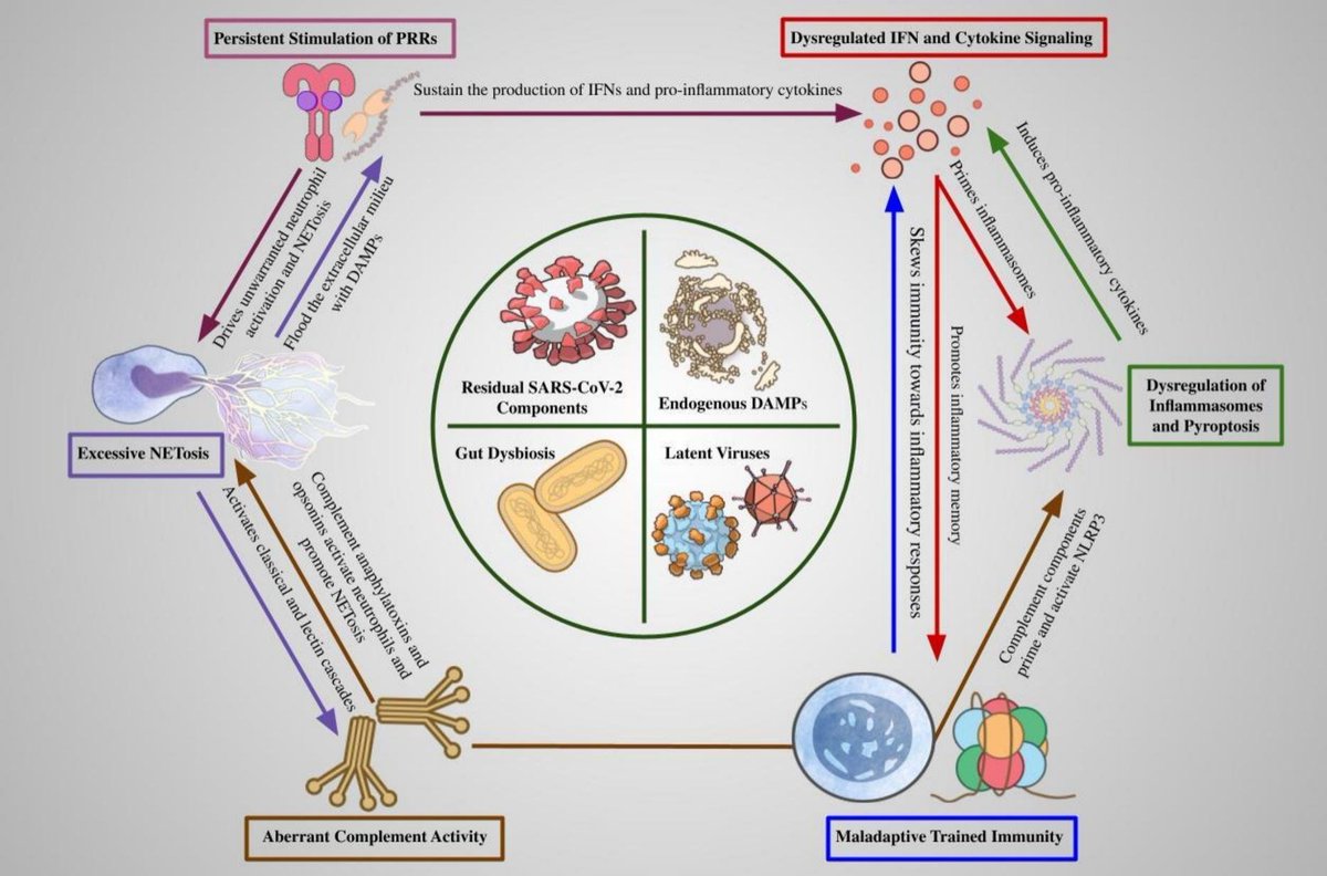
frontiersin.org/journals/immun… transcriptional signatures, LC-ME/CFS patients exhibited a mark⬇️ in naïve CD4+ and CD8+ T cells, regulatory T cells, MAIT cells, γδ T cells, & expansion of effector T cells. NK cells display⬇️ frequency and altered activation-associated transcriptional factors,
consistent with⬇️ cytotoxic potentials. B cells in LC patients exhibited gene expression profiles indicative of heightened activation, while plasma cells revealed a distinct transcriptional subset expressing NK-associated genes. Platelets and low-density neutrophils were expanded and exhibited enrichment of activated-related transcripts.
Monocyte subsets hv transcriptional skewing characterized by⬇️ expression of phagocytosis-associated genes and increased expression of pro-inflammatory cytokine-related genes/pathways. In contrast, idiopathic ME/CFS patients exhibited less pronounced immune alterations at transcriptional level: while T cell activation was evident, there was no reduction in MAIT or NK cells, nor signs of T cell exhaustion.
FOXP3 expression was upregulated, and B cells and platelets demonstrated dysregulated signatures in idiopathic ME/CFS. Galectin-9–TIM-3 interaction as a potential pathway driving γδ and MAIT cell depletion in LC.