A. Joseph Borelli, Jr., MD

29.2K posts

A. Joseph Borelli, Jr., MD banner
A. Joseph Borelli, Jr., MD

A. Joseph Borelli, Jr., MD

@DocBorelli

🩻 Radiologist & MRI clinic owner | Chaired committee writing U.S. MRI accreditation standards | Filmmaker | All tweets IMO | No investment/medical advice


Bluffton, SC Katılım Mayıs 2013
2.3K Takip Edilen3.6K Takipçiler
A. Joseph Borelli, Jr., MD
We provide that article for evidence-based transparency on what to expect, although the protocol I developed greatly exceeds the protocols of the facilities in that meta-analysis: we produce far more images at higher resolution and at 3T. The procedure lasts 90 minutes which is significantly longer and more comprehensive, as well. The article speaks to whole body MRI screening, especially with relevance to detection of neoplasms. The benefits are not yet proven in this regard, mainly because of the limitations of treatments for many cancers. But we believe that will change rapidly with AI advances in pharmacotherapy. We have detected malignant neoplasms in asymptomatic individuals, but we can’t yet prove that earlier detection and treatment led to better outcomes. Still, I’d rather know sooner than later. We detect many silent conditions, some of which are immediately life-threatening, that lead to pharmacologic, surgical, and/or lifestyle interventions that likely extend health-span, including aneurysms, vascular malformations, fatty liver, etc. mriatbelfair.com/uploads/b/ed76…
English
0
0
0
9
Eric Topol
Eric Topol@EricTopol·
The aorta doesn't get enough respect. Even though it's uncommon, in someone who presents with chest pain, especially severe, we were trained to rule out aortic dissection before anything else. Most likely Senator Graham had a Type A dissection and longstanding hypertension. If obtained previously, an echocardiogram or other image may have revealed a widening or aneurysm of the thoracic aorta. nature.com/articles/nrdp2…
Eric Topol tweet media
English
50
207
962
404.1K
A. Joseph Borelli, Jr., MD
Thanks for your inquiry. Read about it here. We've diagnosed many non-vascular lesions, such as severe fatty liver (MASH) and treatable neoplasms as well as many vascular lesions, including a grade 3 DAVF with a 15% annual mortality risk treated with Gamma Knife at Johns Hopkins. mriatbelfair.com/whole-body-scr…
English
1
0
1
17
Rajeev Varma, MD
Rajeev Varma, MD@RVarmaMD·
@DocBorelli @EricTopol You do whole body MRI for screening ? Do you specify it’s strictly for vascular lesions? What’s your protocol ( you can refer to a publication)?
English
1
0
0
27
A. Joseph Borelli, Jr., MD
@SDembraski Apparently not intended for submission to the robotaxi fleet. I don't see customer cars participating in 2026 or 2027 in any meaningful way.
English
0
0
3
166
Scott Dembraski
Scott Dembraski@SDembraski·
Model YL does not have camera washers for front fenders or the rear trunk camera. We were hoping they may start to include them for customer cars. But so far not yet.
Scott Dembraski tweet mediaScott Dembraski tweet media
English
25
5
128
17.3K
Stephan
Stephan@sbinc70·
@DocBorelli @kshpil @EricTopol I’ve had 4 surgeries since. AAA twice, descending repair and tvar for aneurysm at the iliac arteries after descending repair.
English
1
0
0
22
A. Joseph Borelli, Jr., MD
@RVarmaMD @EricTopol That's definitely an acceptable standard of care for follow-up, but not for screening, in my opinion. We need to screen the entire aorta, not just the aortic root and ascending segment. With whole-body MRI, we see the entire thoracic aorta and abdominal aorta.
English
1
0
0
20
Rajeev Varma, MD
Rajeev Varma, MD@RVarmaMD·
@DocBorelli @EricTopol Echocardiogram detects is very well. Plus you can grade any valvular issues. Conservative management is tight blood pressure control.
English
1
0
1
48
Stephan
Stephan@sbinc70·
@DocBorelli @kshpil @EricTopol My problems started with an ascending dissection which damage the aortic valve. Open heart surgery to repair and replace valve in 2003. Later diagnosed with Marfan syndrome by genetic testing at the John Ritter Foundation. I was 31.
English
1
0
0
30
A. Joseph Borelli, Jr., MD
Our procedure does cost more than what many others charge, but it's the equivalent of 8 separate MRI procedures heavily discounted in a package for which most hospitals would charge $25-50K. And it includes consultation with me immediately after the procedure, reviewing results of what we found in about 5,000 images of your entire head, neck, chest, abdomen and pelvis, angiograms of the head and neck and quantitative determination of liver fat. Also, the best MRI scanners alone now cost about $3MM. It was money well spent for our patient with the 6 cm aortic aneurysm missed at a major teaching hospital. We likely saved his live and the lives of many, many others (or at least extended them).
English
0
0
0
7
A. Joseph Borelli, Jr., MD
Having an aneurysm of your ascending aorta increases your risk of dissection. The majority of dissections occur in ascending aortas over 5.0 cm. This is why many advanced centers are now operating on patients with 5 cm ascending aortic aneurysms. Lindsey may have had an aneurysm as the underlying cause. We may never know.
English
1
1
3
235
KAS
KAS@kshpil·
@DocBorelli @EricTopol Different entity than a dissection. Most dissections occur in normal caliber aortas.
English
1
0
0
247
Rod Laver
Rod Laver@rodlaver·
What an exceptional men's final at Wimbledon! Alexander Zverev put up a sterling fight with bold ball-striking, but Sinner rose to every challenge. Congratulations to Jannik on another Wimbledon title. 🚀
English
26
132
1.8K
25.3K
BRICK BY BRICK
BRICK BY BRICK@aincomeinvestor·
I will never delete reddit.
BRICK BY BRICK tweet media
English
147
285
54.6K
3.6M
👀
👀@Flavio_M24·
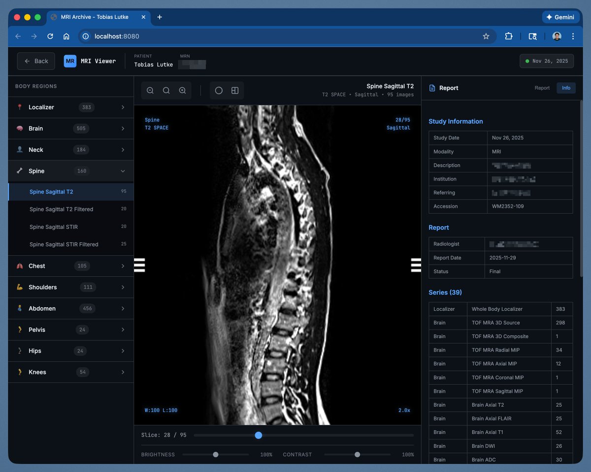
From the image you shared (sagittal T2 SPACE of the spine, slice 28/95, likely mid-thoracic or lower thoraco-lumbar region), we can clearly see the posterior aspect of the spine including vertebral bodies, discs, spinal canal, and cord/thecal sac. The most noticeable finding (and quite common in this kind of imaging) is **mildly increased (hyperintense) signal in the central or central-posterior portion of the spinal cord** on T2, particularly in the visible thoracic segment. This kind of T2 hyperintensity in the cord on a sagittal view can correspond to several possibilities (listed roughly in order of likelihood in routine imaging): 1. **Flow-related artifact or CSF pulsation artifact** → very common in T2 SPACE (3D sequence), especially mid-to-lower thoracic levels. The cord often looks “dirty” or shows central ill-defined hyperintensity that is not a true lesion. 2. **Mild/atypical degenerative myelopathy / chronic compression-related signal change** → if there is significant canal stenosis or large disc herniations higher up (not well seen in this single slice), you can sometimes get focal or “snake-like” T2 hyperintensity in the cord. 3. **Low-grade syrinx / hydromyelia** → if the hyperintensity is very well-defined, central, and tubular → but in this image it doesn’t look particularly convincing. 4. **Sequelae of myelitis / demyelination** (e.g., post-viral, MS-like, neuromyelitis optica, B12 deficiency) → usually more extensive and/or with different patterns (posterior, lateral, “inverted V”, bright spotty lesions…). 5. **Chronic spinal cord ischemia / vasculopathy** → typically more segmental and associated with suggestive clinical history. **What’s missing to better understand it?** Pretty much everything else 😅 - Axial images at the same level (to see if it’s central, posterior, lateral, occupies >2/3 of the cord, enhancement…) - The STIR series (already present in your archive) → much more sensitive for true cord edema - The filtered series you mentioned (“Spine Sagittal T2 Filtered” / “STIR Filtered”) → often used to reduce artifacts and better evaluate the cord - Exact level (C7-T1? T8-T9? T11-L1?) and whether it continues in adjacent slices - Any obvious mechanical compression (stenosis, herniation, listhesis…) - Age, symptoms, and the official radiology report **Short conclusion** In this single image, the most likely thing that stands out is a **flow/artifact-related appearance in the thoracic cord** (common and usually benign in T2 3D SPACE sequences), or a **mild cord T2 hyperintensity of uncertain clinical significance** (should be compared with STIR and axials). If you have cord-related symptoms (spasticity, sensory changes in the legs, sensory bands, urgency/incontinence…), then it should be taken seriously and correlated with the full exam. Otherwise, it’s often an “incidentaloma” or just an artifact.
English
1
0
0
74
tobi lutke
tobi lutke@tobi·
My annual MRI scan gives me a USB stick with the data, but you need this commercial windows software to open it. Ran Claude on the stick and asked it to make me a html based viewer tool. This looks... way better.
tobi lutke tweet media
English
1K
1.8K
33.9K
8.5M
A. Joseph Borelli, Jr., MD
Yeah. So why do you think he's going to put AI5 into cars any time soon when last year he said AI5 was coming to cars but at the Q1 earnings call this year he reneged, saying AI5 was destined for Optimus and Cortex (training)? The next chip upgrade for cars is AI4 Plus with doubled RAM and a slight performance boost, but not AI5.
English
0
0
0
22
Whole Mars Catalog
Whole Mars Catalog@wholemars·
AI5 will be 2 nanometers and has reached tape out!
Sawyer Merritt@SawyerMerritt

Samsung Foundry’s Principle Engineer has announced that “the @Tesla-Samsung Al5 chip has reached tape-out. It is scheduled to be manufactured at the Taylor fab using our latest 2nm process and will soon be integrated into Tesla's newest products.” Volume production won’t start for a while, but good to hear things are still moving along. Picture of Tesla’s upcoming AI5 chip on the right:

English
32
38
787
62.5K
Cult Buster
Cult Buster@PabluGablu·
@DocBorelli @wholemars Wrong. 😑 Sorry, but AI4 will be outdated sooner than you think. Saying AI4 is the final answer is like claiming Apple would stop at the iPhone 11 and never build the 12, 13, 14, 15, or 16. First lesson: learn how technology works. It evolves.
English
1
0
1
29
A. Joseph Borelli, Jr., MD
Accurate. Elon’s recent statements: In the Q1 2026 earnings call, Elon Musk said AI5 is expected to go into Optimus and data centers because unsupervised self-driving can be achieved with AI4, directly contradicting the claim that he said it would go into Tesla cars as well. • Plans evolved from earlier comments: In a January 2026 X post, Musk noted AI5 would “make the cars almost perfect and greatly enhance Optimus,” but later updates including the earnings call and April tape-out announcements prioritized AI5 for robots and compute clusters first.
English
0
1
1
141
Overly Trev
Overly Trev@OverlyTrev·
No. Elon said they’re not putting AI5 in production cars. Did anyone listen to the Q1 earnings call? lol. Also making a Model for AI5 would be a complete waste of time right now. 99% of the fleet wouldn’t be able to run the model at the beginning, it would have to be distilled down. AI5 is for Optimus and for inference compute. AI4 is already unsupervised and we are getting a larger model with V15 on AI4. I bet AI5 won’t be in customer vehicles until 2028 or later.
Dirty Tesla@DirtyTesLa

Model YL HW5 next year? 👀

English
39
5
206
33.5K
A. Joseph Borelli, Jr., MD
No, the post is not accurate on Elon’s recent statements: In the Q1 2026 earnings call, Elon Musk said AI5 is expected to go into Optimus and data centers because unsupervised self-driving can be achieved with AI4, directly contradicting the claim that he said it would go into Tesla cars as well. • Plans evolved from earlier comments: In a January 2026 X post, Musk noted AI5 would “make the cars almost perfect and greatly enhance Optimus,” but later updates including the earnings call and April tape-out announcements prioritized AI5 for robots and compute clusters first.
English
1
0
0
59