ECG Reid
557 posts


ECG Reid
@ECGwithReid
#ECG Education for Physician Assistants & Nurse Practitioners
Katılım Ekim 2020
200 Takip Edilen2.4K Takipçiler

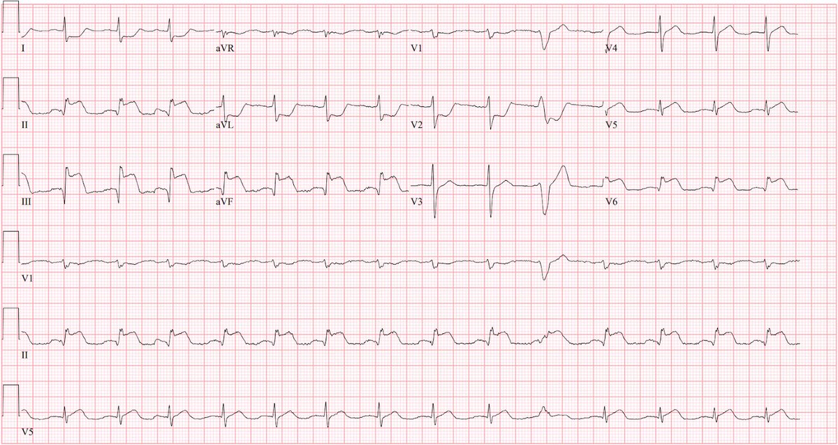
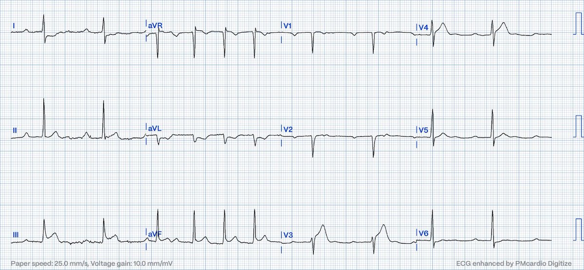
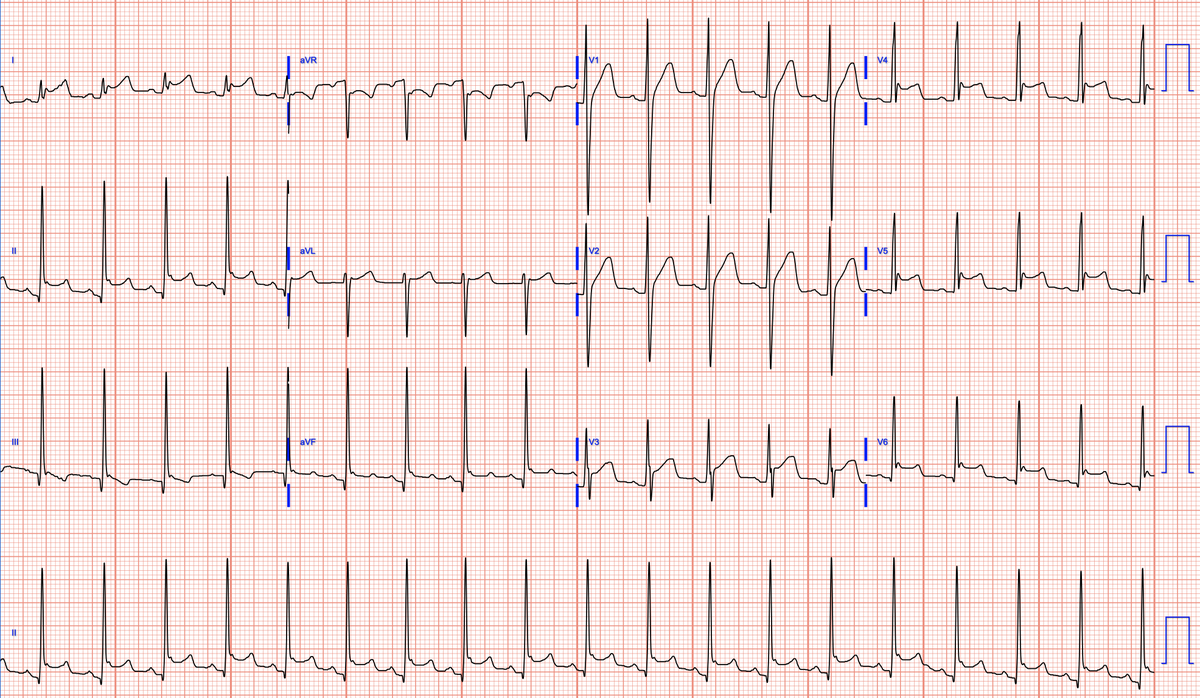
This ECG was sent to me with the question: SVT with RBBB aberration or VT?
@EM_RESUS @UlhasDr @The_Nanashi_O @DidlakeDW @ecgrhythms @AThomazAndrade @willyhfrick @DaveRichley @Carlos_Penate6 @ECGOxford
@syamkumarmd @Arron_Pearce_ @DrRazi4
@drbasitmasoodi @FloydECGs

English
@jalosmar @PendellM @Arron_Pearce_ @anais_dahoumane @ecgandrhythmRoe @EM_RESUS @smithECGBlog @AntoOhanian @BrooksWalsh @drbasitmasoodi @ECGEPSCADEVICE @ecgotaku @EcgOxford @EcgsOnly Smells like Pericarditis, but I have been fooled many of times!
English

Male, 51 YO. CP since 1h ago. @PendellM @Arron_Pearce_ @anais_dahoumane @ecgandrhythmRoe @EM_RESUS @smithECGBlog @AntoOhanian @BrooksWalsh @drbasitmasoodi @ECGEPSCADEVICE @ecgotaku @EcgOxford @ECGwithReid @EcgsOnly
HT
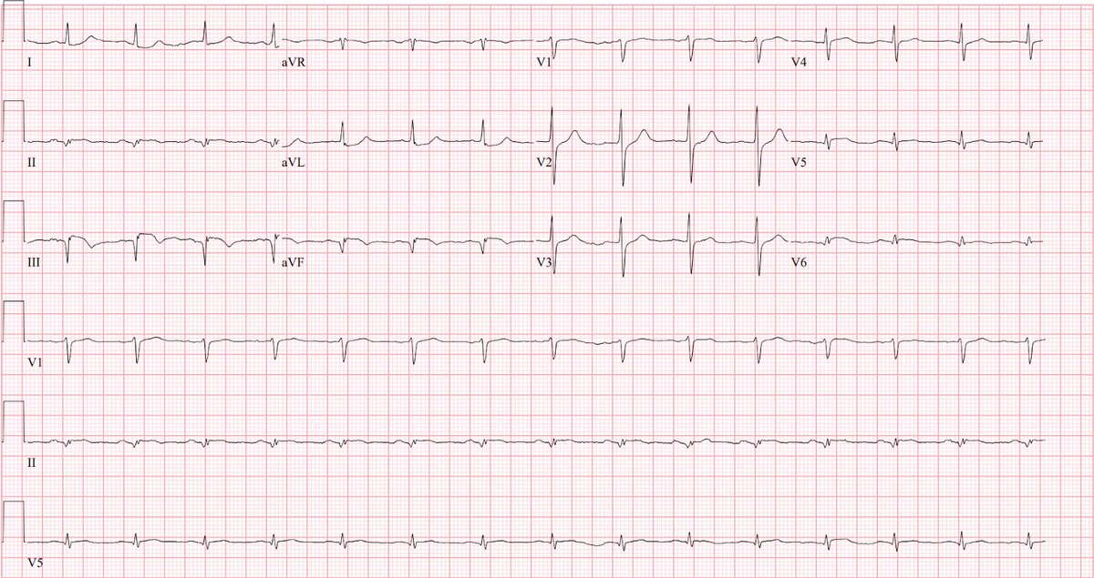
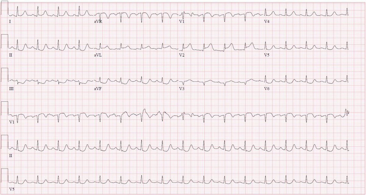
@ECGwithReid Narrow complex tachycardia; what is the answer @ECGwithReid ?
English
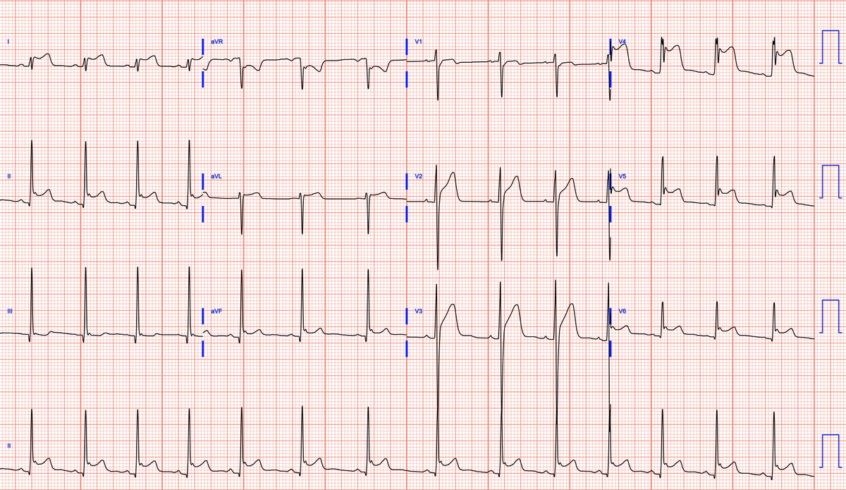
@heartsECGcourse @ECGcases @EMCases RCA occlusion until proven otherwise! Inferior hyper acute T waves, posterior involvement, complete heart block! High lateral reciprocal changes
English

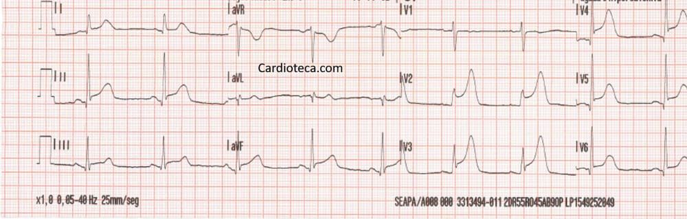
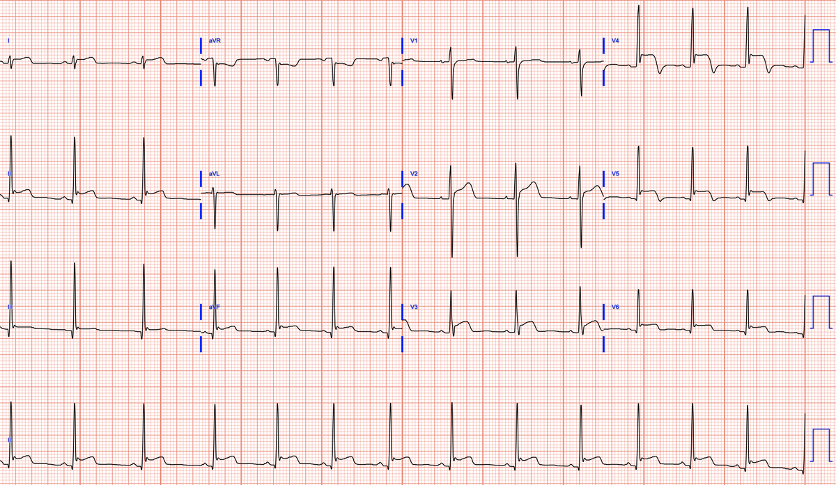
60 year old with epigastric pain and nausea, STEMI negative. What do you think?
@ECGcases @EMCases
#ECG #EKG #ECGinterpretation #foamed #MedEd #medstudent #resident #nurse #paramedic #emergencymedicine #CardioTwitter

English
@willyhfrick @EM_RESUS @BrooksWalsh @FloydECGs @DidlakeDW @The_Nanashi_O @EcgsOnly @RobertHermanMD @DrRazi4 @Vadeboncoeur_Al @DrparrayMD RCA culprit here. I think I see sinus tachycardia with intermittent 2:1 AV block (best seen in V2) 🤔
English

ECG Reid retweetledi
ECG Reid retweetledi
ECG Reid retweetledi

Chest pain and ST elevation, in sequence
@RobertHermanMD @smithECGBlog @EM_RESUS @BrooksWalsh @DidlakeDW

English
ECG Reid retweetledi

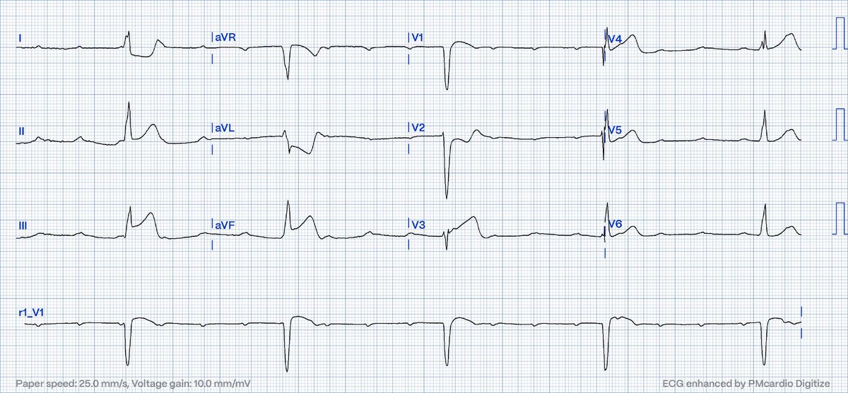
Differentiation of WCT…,
〈Brugada Algorithm〉
・No RS in V1-6
➥VT is likely
〈Vereckei aVR Algorithm〉
・Initial q wave>40 msec
➥VT is likely
+"Positive concordance" in chest leads
▷▷▷VT is likely, I think.
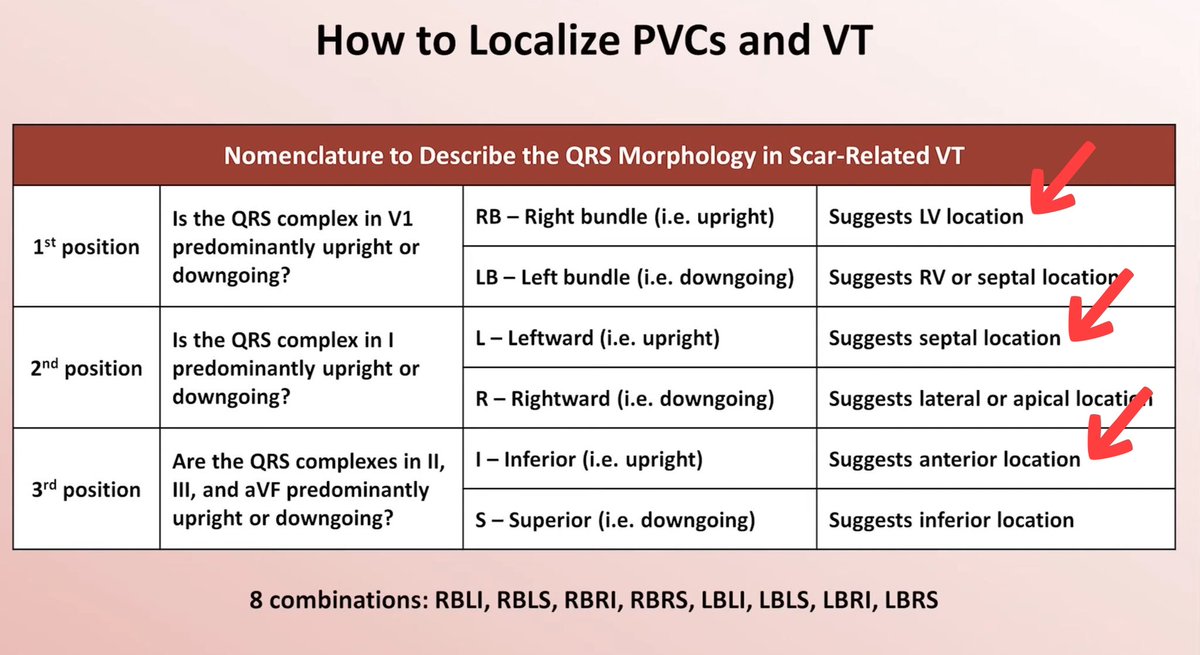
Using this figure, the origin of this VT is "RBLI pattern"
➥Antero-septal wall side of LV origin🧐🧐🧐
English
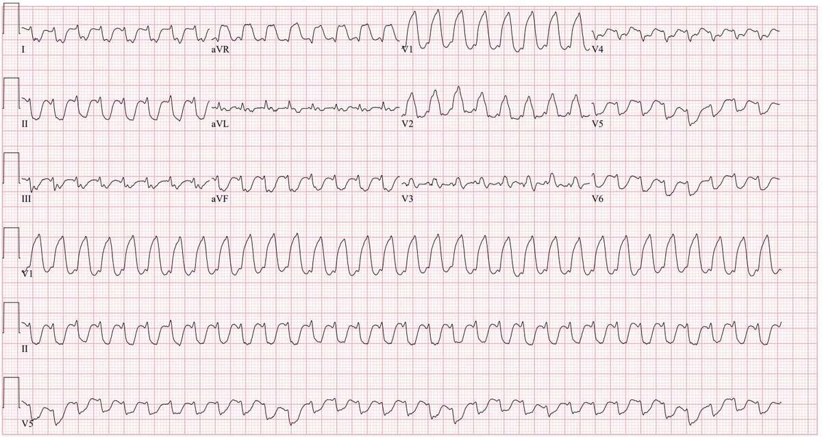
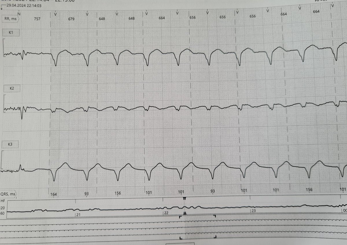
@IrmaMDv Personally, I don't think there are two different competing atrial and ventricular rhythms.
Can see similar WRS widening in some of the PAC's. Post tachycardia pause. I vote some sort of SVT/Atrial-based tachycardia with aberrancy. Open to other opinions 😀!
English


@heartsECGcourse Likely warrants provocative testing for Brugada.
English
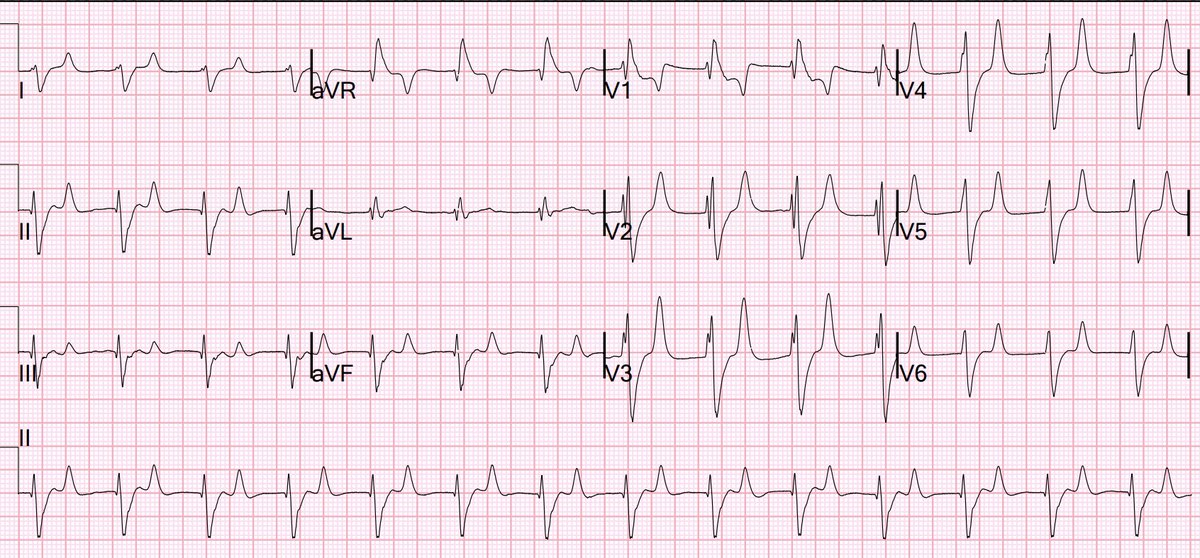
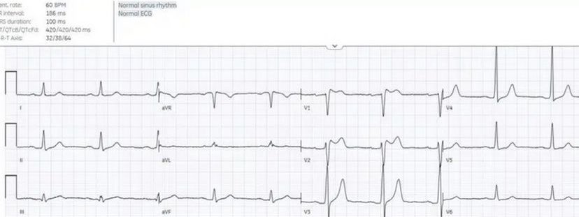
50 year old with syncope. Is this ECG normal?
#ECG #EKG #FOAMed #CardioEd #CardioTwitter #medstudent #MedstudentTwitter #paramedic #EmergencyMedicine
English
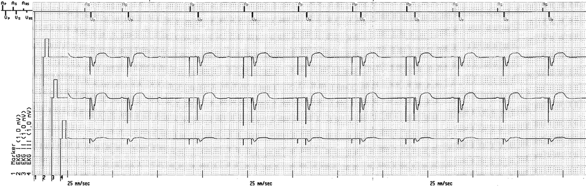
@ecgandrhythmRoe Looks like a few episodes of unsensed atrial depolarization!
English
@ECGwithReid Maybe a type 2 myocardial infarction; the RCA is not severely narrowed or occluded, but the drop in Hb has nevertheless triggered an IWMI with posterior involvement?
English
Interesting case. 65 year old male with chest pain. Found to have splenic rupture on imaging. Taken to the OR for splenectomy. 2L blood loss. Initial Hgb 10 but down to 7 g/dL postoperatively. However, on chart review, initial troponins continued to rise over next few days. Finally, ECG obtained. Thoughts? Demand ischemia or occlusion?
English
Another interesting case. 60 year old female with history of IgG lambda myeloma presented to the ER in SVT. Found to have pericardial effusion with suspicious LV mass. Pericardial fluid positive for lambda light chain restricted population of plasma cells.
Diagnosis: plasmacytoma causing malignant pericardial effusion.
English
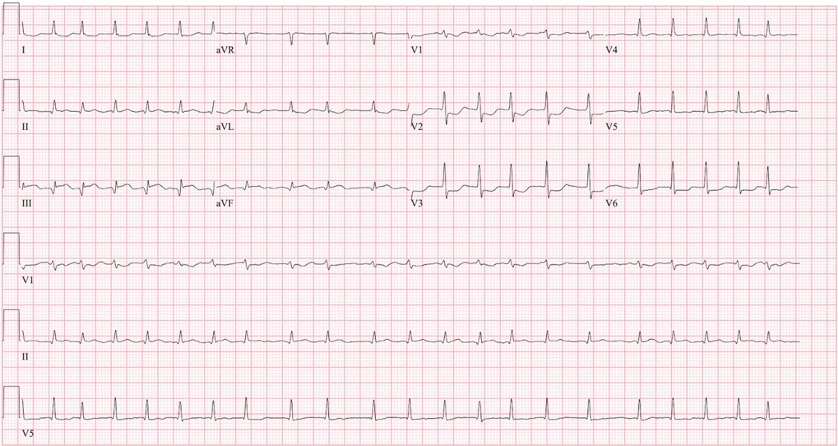
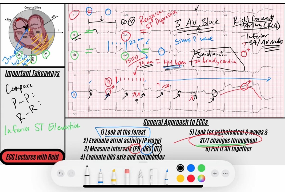
Here I break down an #ECG with a Right Coronary Artery (RCA) occlusion and Complete AV Block 😁
Link to YouTube ⤵️
youtu.be/Nk-i2BRO0OU?si…

YouTube
English