AANP

1.8K posts

AANP banner
AANP

AANP

@neuropathology

The official X account of the American Association of Neuropathologists (AANP)

Littleton, CO Katılım Ocak 2009
840 Takip Edilen4K Takipçiler
AANP
AANP@neuropathology·
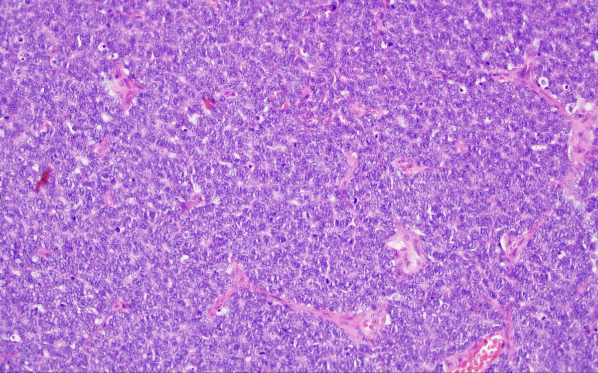
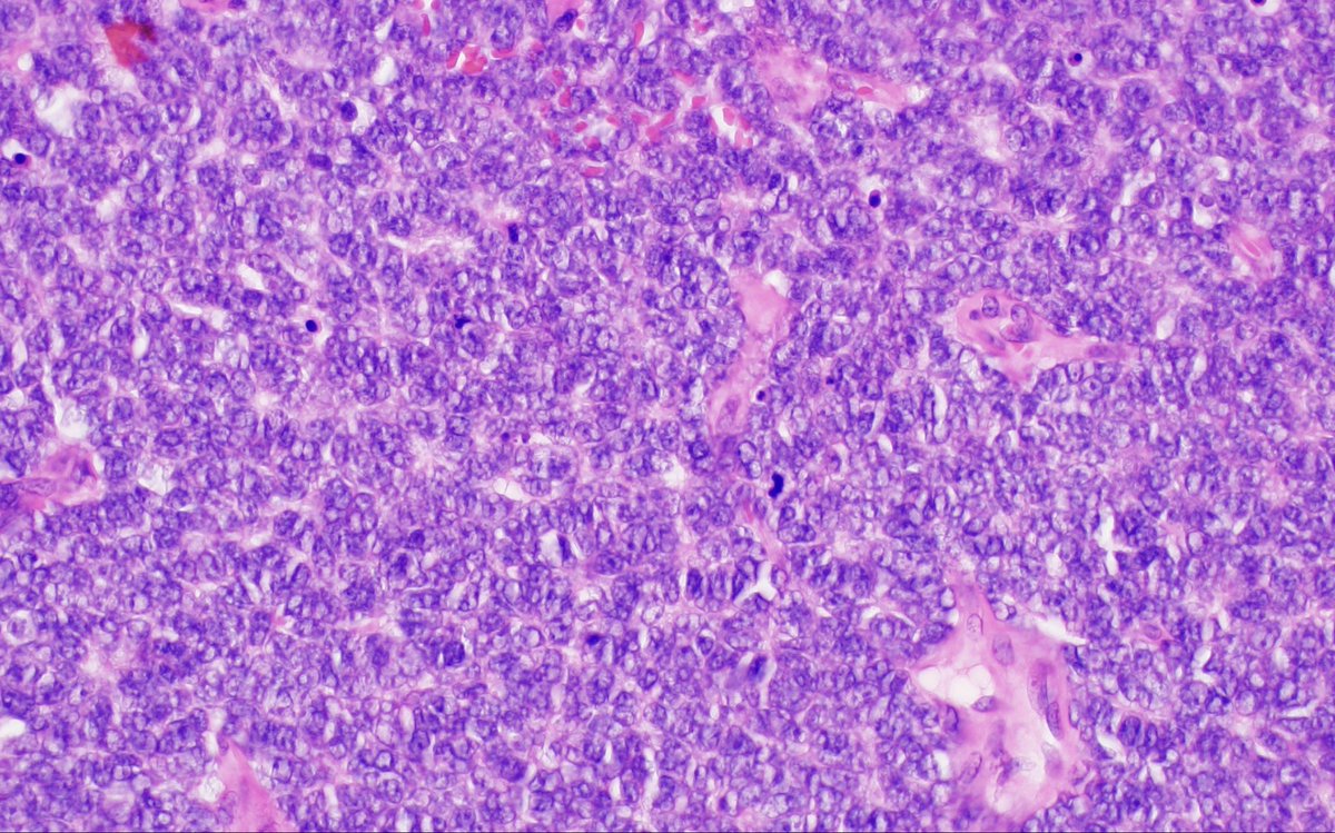
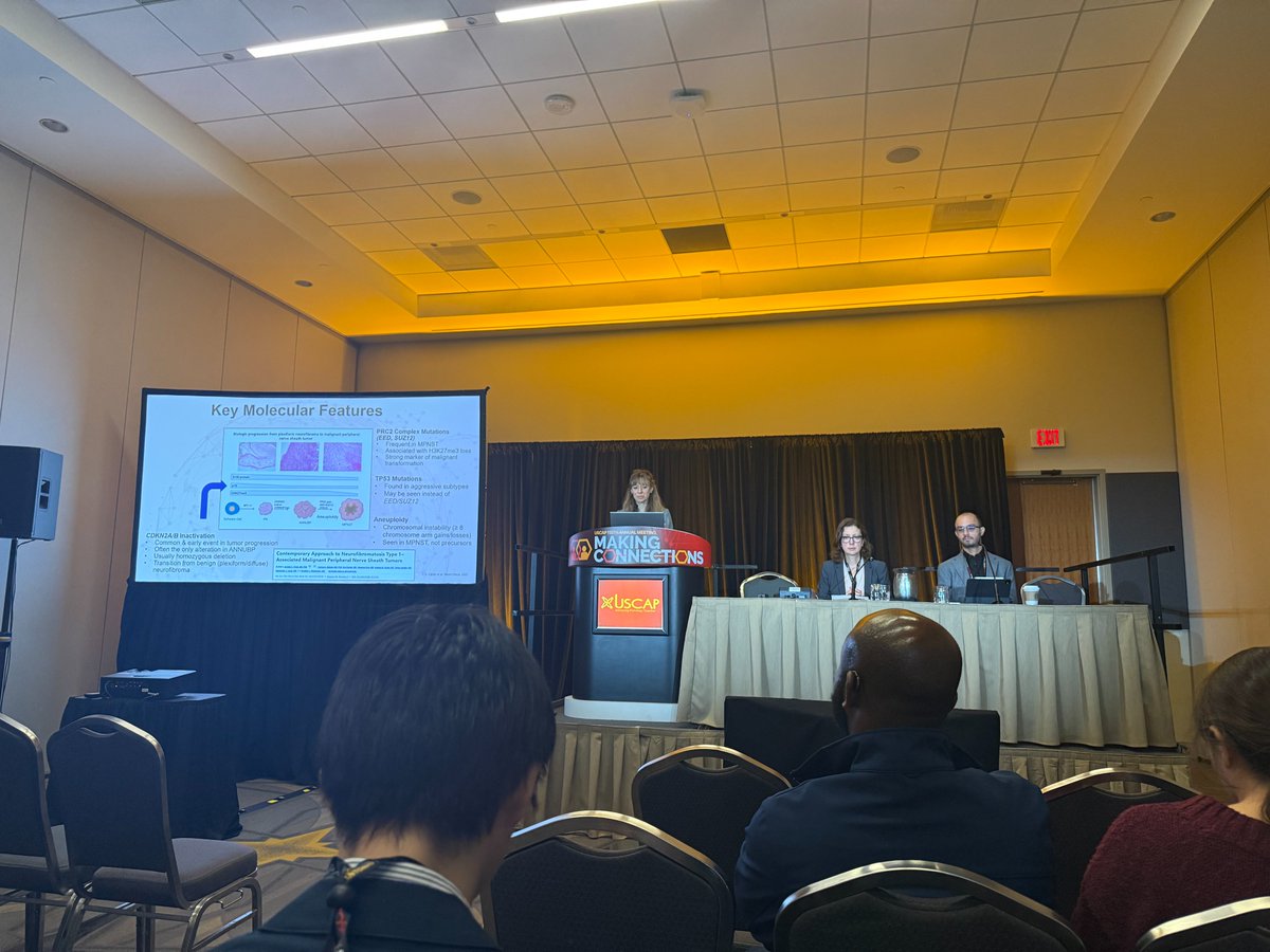
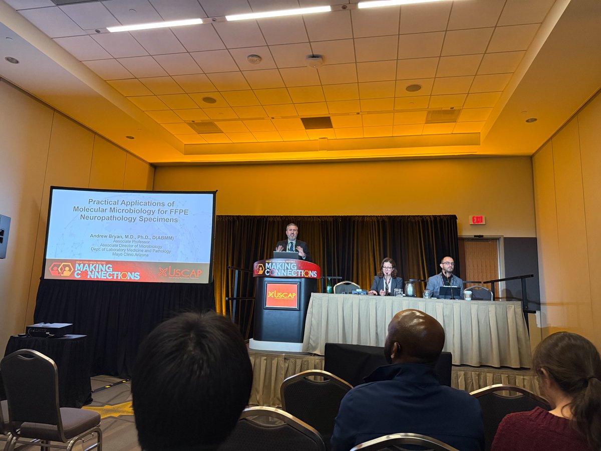
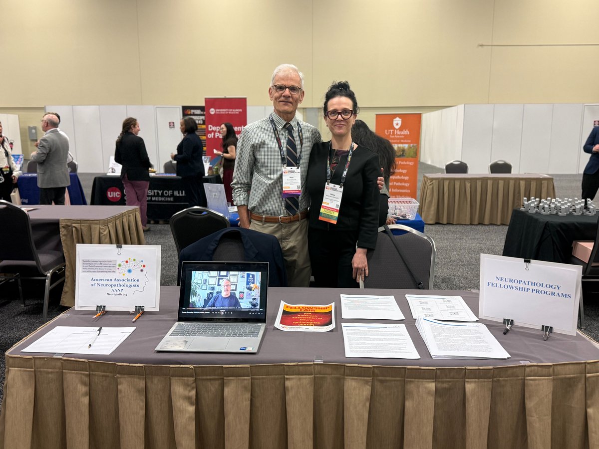
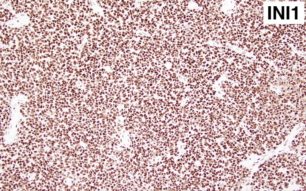
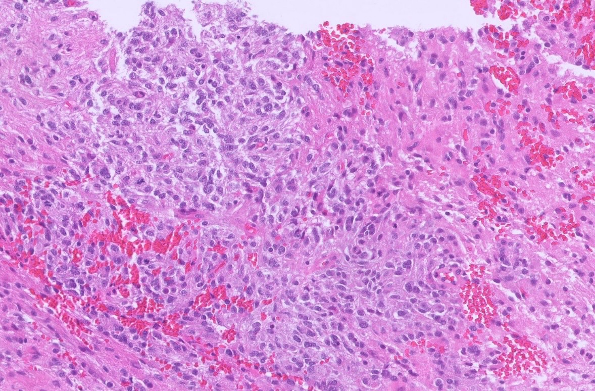
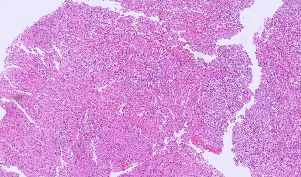
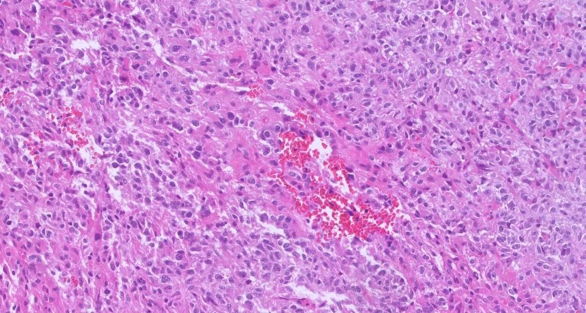
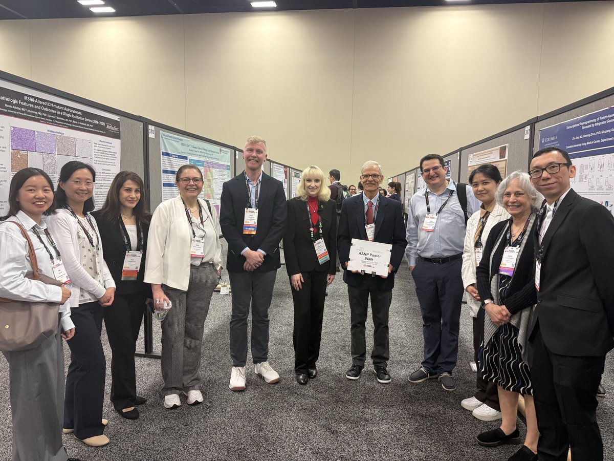
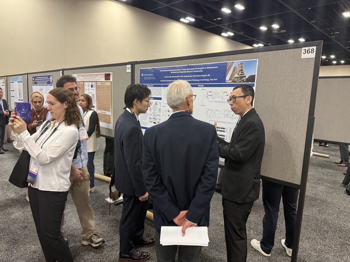
The @neuropathology poster walk at #USCAP2026 was a great success! It was wonderful to see all of the #neuropath representation and connect with colleagues.
AANP tweet mediaAANP tweet mediaAANP tweet mediaAANP tweet media
English
0
6
28
1.5K
AANP
AANP@neuropathology·
The correct diagnosis is Ewing sarcoma, which was confirmed by detection of the EWSR1::FLI1 fusion transcript. Please visit the AANP website for full case discussion! aanp.memberclicks.net
English
0
0
0
75
AANP
AANP@neuropathology·
⭐️AANP Case of the Month⭐️ Credit: @JaredAhrendsen 5yo p/w large, variably enhancing frontal mass. There is no evidence of extracranial disease. Resection reveals histomorphology shown here. What is your differential diagnosis? Which IHC/stains would you order? #neuropath
AANP tweet mediaAANP tweet media
English
5
8
27
2.5K
AANP
AANP@neuropathology·
We had a wonderful AANP Companion Society Meeting this morning at #USCAP2026! The Fellowship Fair was another huge success! Don't forget to join us again tomorrow Monday 3/23 at 9:45am for a Poster Walk starting at poster #366!
AANP tweet mediaAANP tweet mediaAANP tweet mediaAANP tweet media
English
0
2
10
723
AANP
AANP@neuropathology·
The American Association of Neuropathologists will be represented at #USCAP2026, March 21-26, 2026, in San Antonio, Texas, through the following events. If you’re planning to attend, please join!
AANP tweet media
English
0
3
7
794
AANP
AANP@neuropathology·
The differential for this malignant small round blue cell tumor is broad, including CNS embryonal neoplasms and poorly differentiated sarcomas. Representative IHCs are shown. What is the most likely diagnosis? What molecular finding(s) do you expect to be present in this tumor?
AANP tweet mediaAANP tweet mediaAANP tweet media
English
2
4
7
773
AANP
AANP@neuropathology·
HGG-F (Adult-type diffuse high-grade glioma, IDH-subtype F) an emerging new type of high-grade glioma, often with a gliomatosis-like pattern. Interestingly, they seem to have a better prognosis compared to Glioblastoma. #neuropath #neuropathology #pathx #PathTwitter
AANP tweet mediaAANP tweet mediaAANP tweet media
English
0
6
21
1.1K
AANP
AANP@neuropathology·
The ddx is broad: meningioma, solitary fibrous tumor, other CNS mesenchymal lesions, and metastatic sarcoma. Useful IHC/stains shown here suggest a CNS mesenchymal tumor. Final diagnosis of intracranial mesenchymal tumor, FET::CREB fusion-positive, was confirmed by molecular.
AANP tweet mediaAANP tweet media
English
1
0
1
217
AANP
AANP@neuropathology·
⭐️AANP Case of the Month⭐️ Credit: Dr. Purvi Patel, MD, PhD 66F p/w seizure. MRI: left frontal extra axial mass. Based on the images shown, what is your DDX? What stains would you order? Likely molecular alteration? #neuropath
AANP tweet mediaAANP tweet mediaAANP tweet mediaAANP tweet media
English
3
9
39
3.5K
Charlie Herndon
Charlie Herndon@DrCycloPath·
🔬 #CytoQuiz #GUPath A) Chromophobe Renal Cell Carcinoma B) Clear Cell Renal Cell Carcinoma C) Papillary Renal Cell Carcinoma D) Adrenocortical Carcinoma
Charlie Herndon tweet media
Català
4
6
27
2.4K
AANP
AANP@neuropathology·
Be sure to check out the AANP website for additional education and neuropathology related content, including previous “Case of the Month” reports! neuropath.org/case-of-the-mo…
English
0
0
3
486
AANP
AANP@neuropathology·
Make sure to visit the AANP website for additional educational content, including full write ups for this and prior months’ posts! neuropath.org/case-of-the-mo…
English
0
0
0
130
AANP
AANP@neuropathology·
⭐️AANP Case of the Month⭐️ Credit: Dr. Dimitri Trembath, MD, PhD 52F p/w worsening HA, visual disturbance, and ataxia. MRI brain reveals an enhancing extra axial mass at the CP angle. Histology shown. DDX? Useful IHC? Likely molecular alterations? #neuropath
AANP tweet mediaAANP tweet media
English
7
15
34
3.1K
AANP
AANP@neuropathology·
This is a chordoid meningioma, with a differential dx including chordoma (esp at/near the skull base). Meningioma markers (EMA+, PR+) help to differentiate from chordoma (bracyury+, S100+)
English
0
0
0
104