Rashna Meunier, MD

5.7K posts

Rashna Meunier, MD banner
Rashna Meunier, MD

Rashna Meunier, MD

@RMeunierMD

Pathologist in upstate NY. Interests: surgical pathology, GI pathology

New York, USA Katılım Ekim 2014
1.2K Takip Edilen3.5K Takipçiler
Rashna Meunier, MD retweetledi
Ziad El-Zaatari
Ziad El-Zaatari@ziad_zaatari·
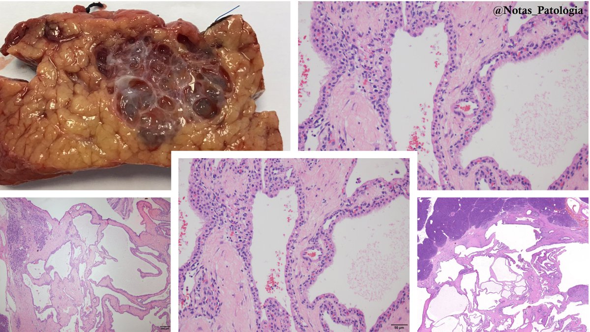
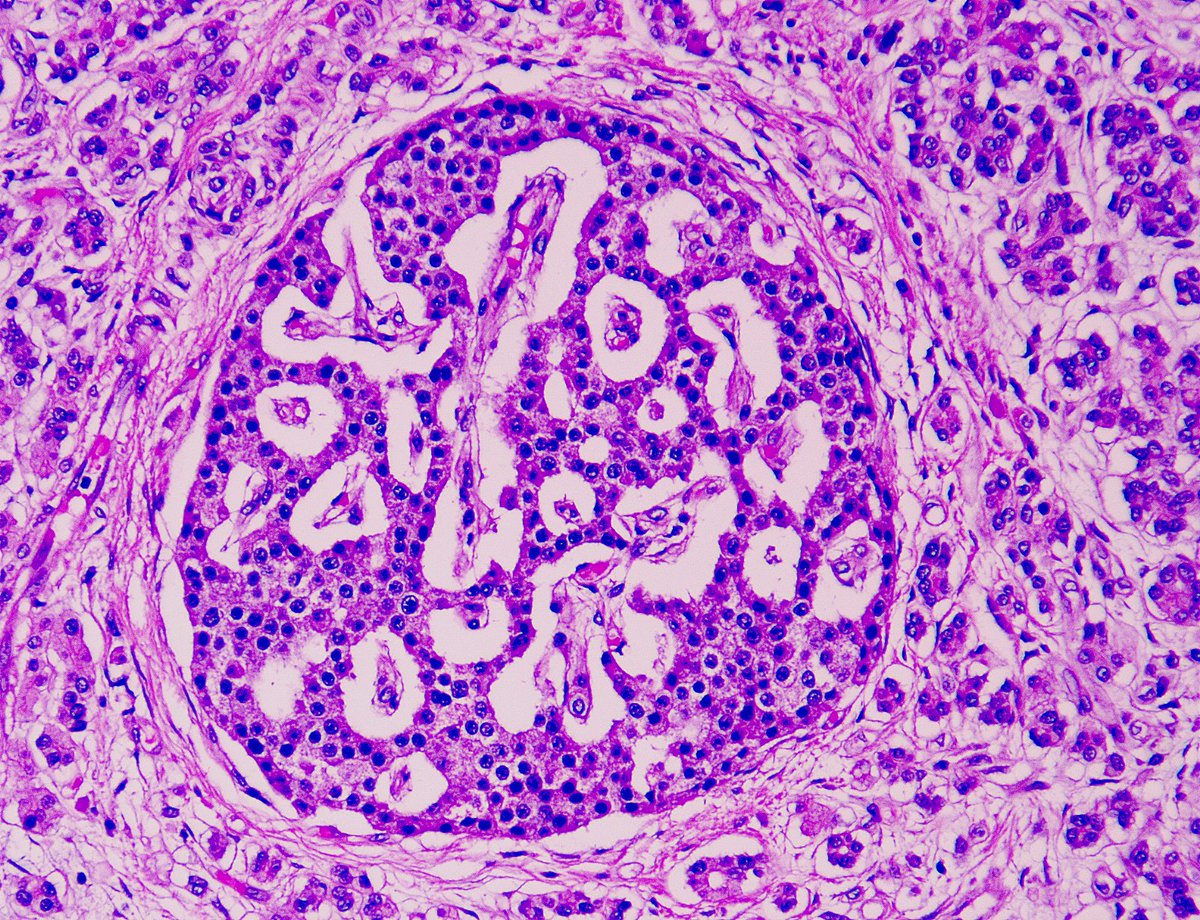
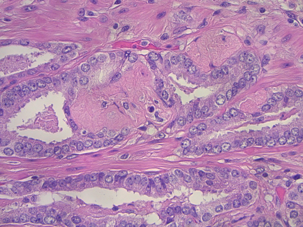
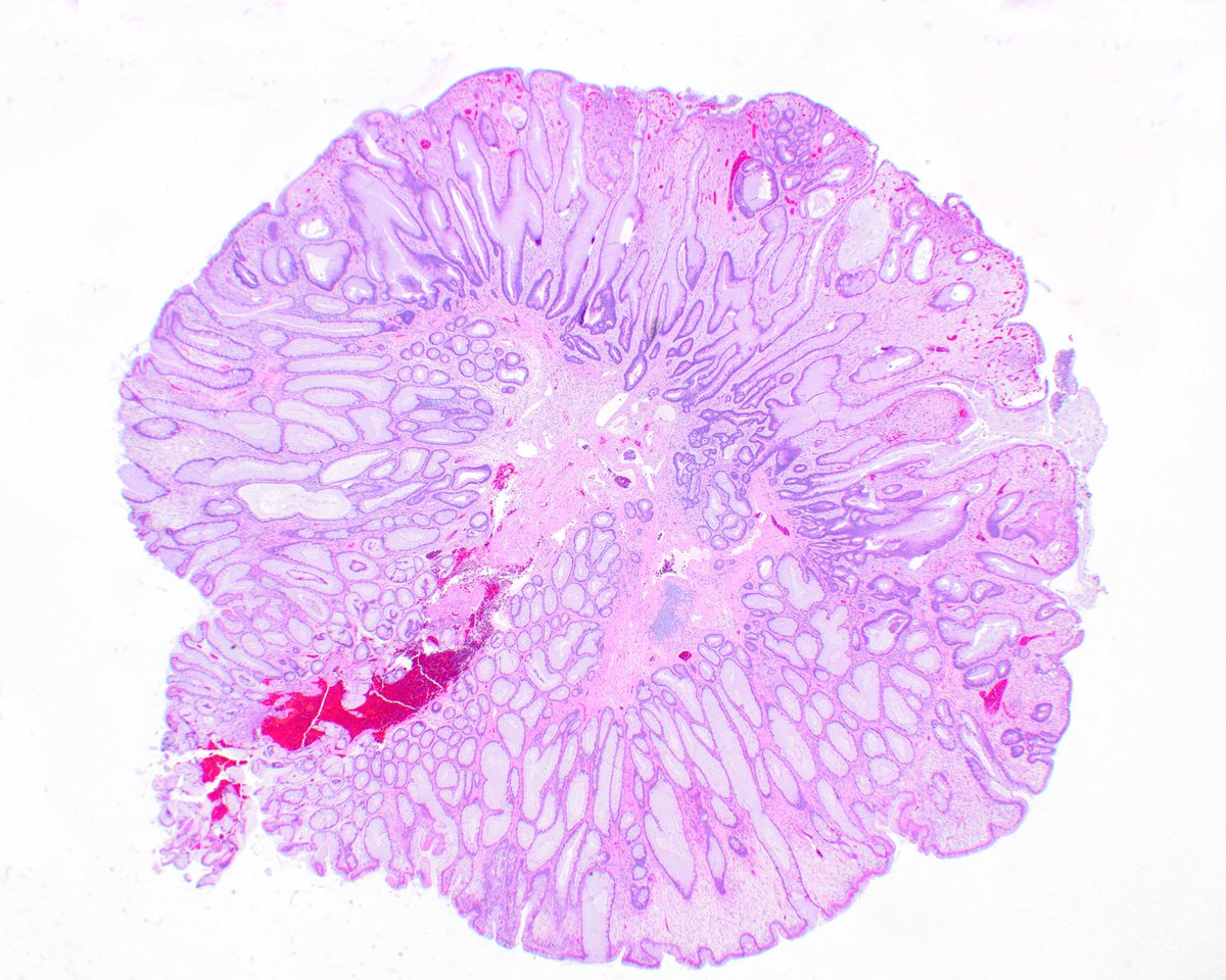
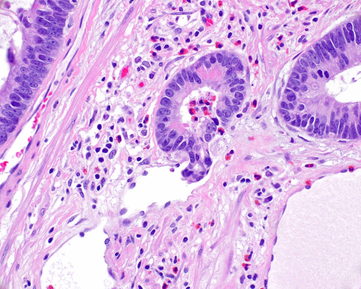
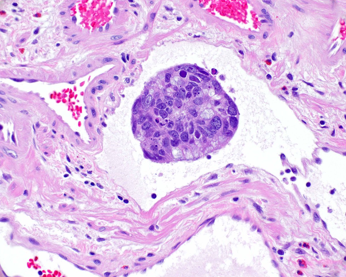
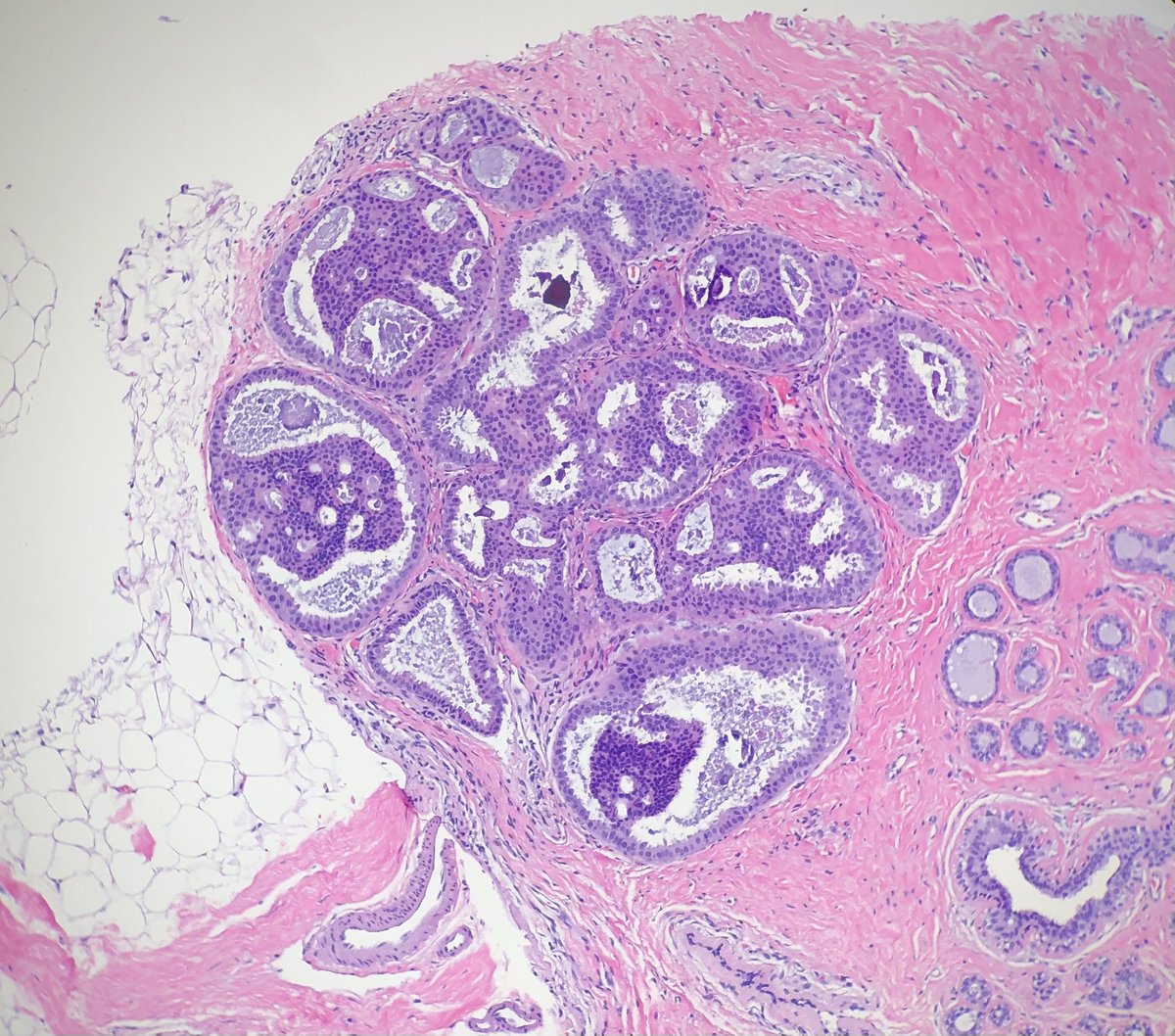
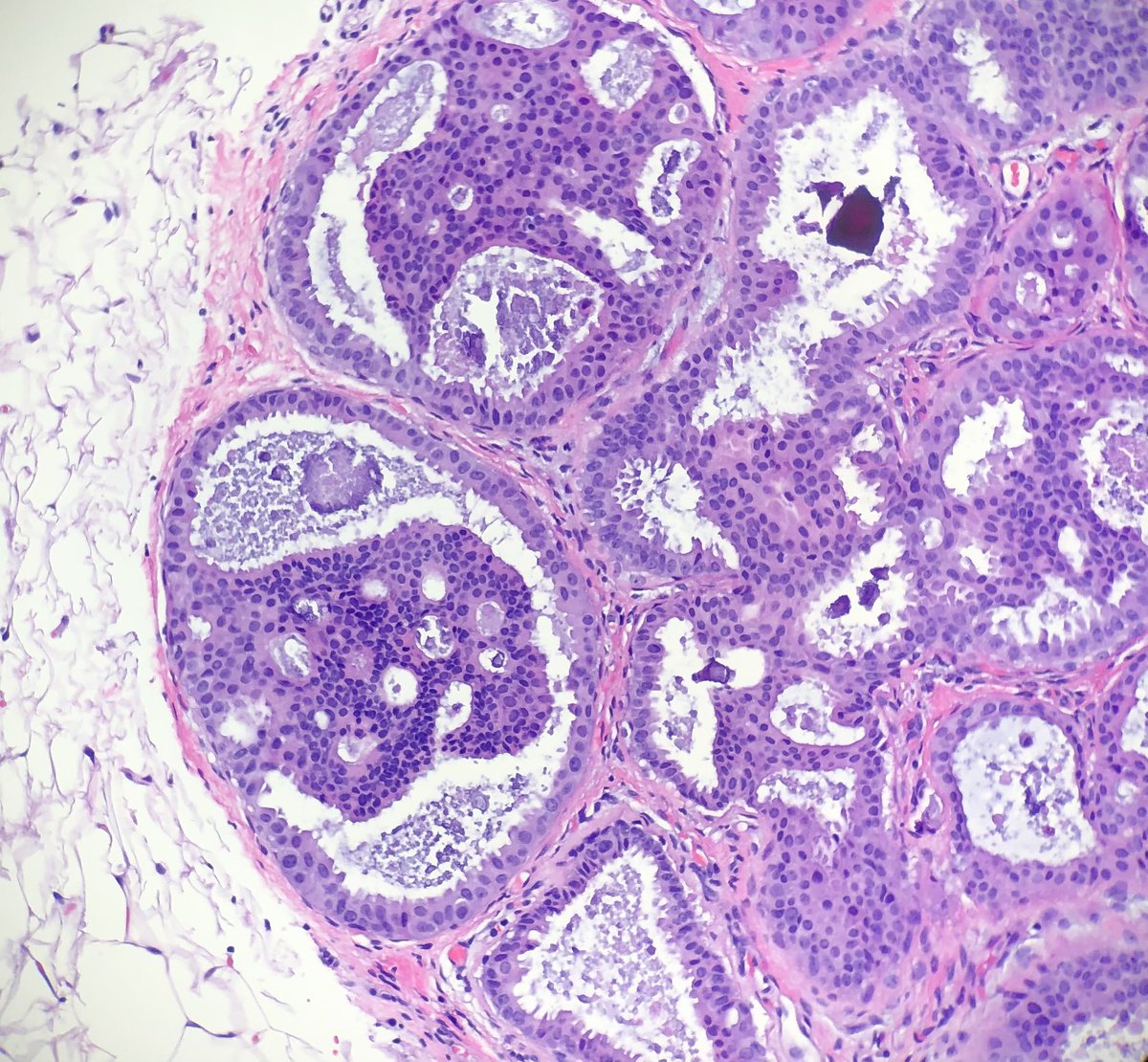
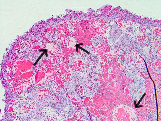
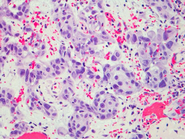
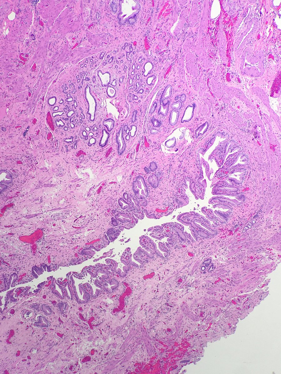
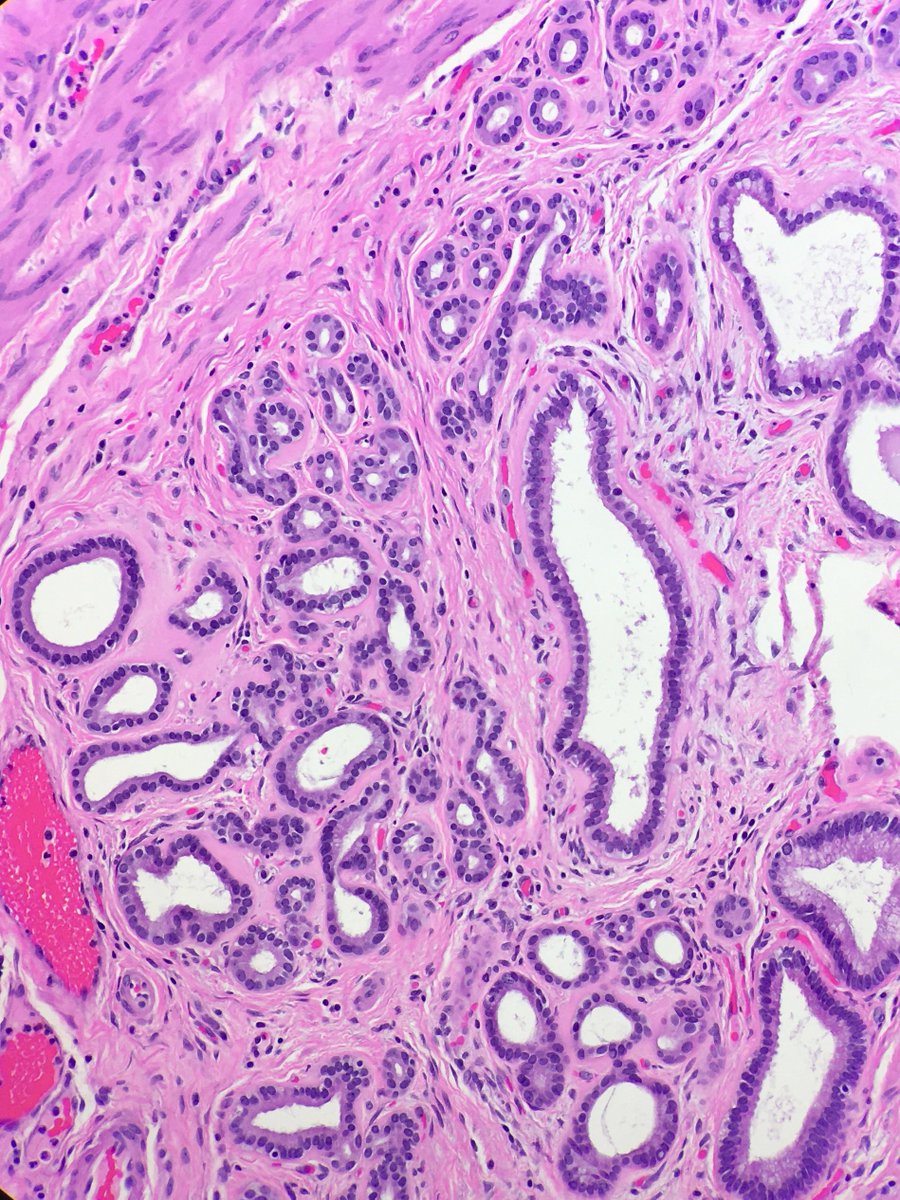
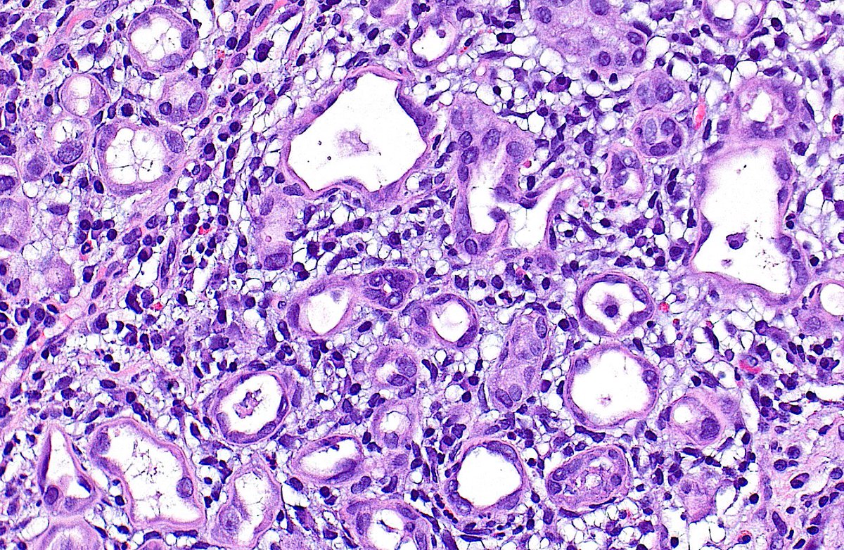
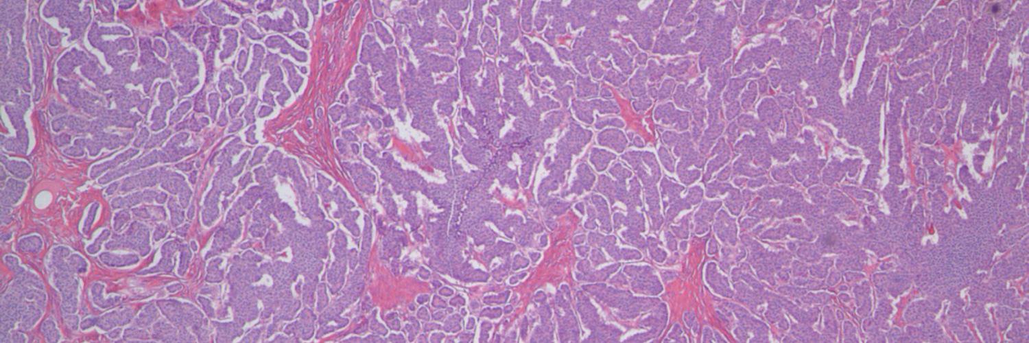
🔬 Nephrogenic Adenoma ~ Tubules lined by single layer of flattened or hobnailed epithelium ~ #GUpath #Pathology #Urology
Ziad El-Zaatari tweet media
English
0
25
99
3.3K
Rashna Meunier, MD retweetledi
Joe
Joe@MBBS_Pathology·
Not always easy to recognise #FlatEpithelialAtypia, but hope this composite image may help. On ⬅️ columnar cell change (elongated bland nuclei). On ➡️ FEA - smooth contour, >1 layer, low grade round nuclei (like LGDCIS), not perpendicular to basement membrane. #pathology
Joe tweet media
English
3
48
194
8.4K
Rashna Meunier, MD retweetledi
NotasDePatologia
NotasDePatologia@Notas_Patologia·
🔬 Serous Cystadenoma of the Pancreas: What You Need to Know Serous Cystadenoma of the Pancreas, also known as Microcystic Serous Adenoma, is a rare benign cystic neoplasm that shows a female predominance (3:1), typically diagnosed around the age of 58. It accounts for about one-third of primary pancreatic cysts. 🧬 In some cases, it may be associated with von Hippel-Lindau (VHL) syndrome, particularly when multifocal. 📍 Anatomical Location: Most frequently found in the tail of the pancreas, with a body/tail to head/neck ratio of about 1.6:1. In VHL-associated cases, it may involve the entire pancreas. 🔍 Gross Features: These are usually well-circumscribed lesions, averaging 6 cm in size but ranging from 1 to 30 cm. The most common type is microcystic, with a sponge-like appearance composed of numerous small cysts filled with clear fluid. Other types include: Oligocystic (Macrocystic): fewer, larger cysts Solid variant: homogeneous, gelatinous, and yellow-beige in appearance A classic feature is the presence of a central stellate scar, consisting of hyalinized fibrous tissue. Hemorrhage may occasionally occur. Multifocality is rare but more common in VHL patients. 🔬 Microscopic Findings: Cysts are lined by cuboidal to low columnar epithelium with clear cytoplasm rich in glycogen (PAS-positive, diastase-sensitive). Nuclei are round, uniform, with dense chromatin, inconspicuous nucleoli, and no mitoses. The stroma may range from edematous to fibrous, sometimes myxoid or hyalinized, and features a rich capillary network. Calcifications can be present. Variants include tiny papillary projections (without fibrovascular cores), oncocytic types with granular cytoplasm, and a solid variant with compact small glands and minimal lumina. 🧪 Immunohistochemistry: Tumor cells are typically positive for EMA, MUC1, pancytokeratin, inhibin, and GLUT1, and negative for HMB45. These markers help distinguish it from other cystic neoplasms. 🧬 Molecular and Cytogenetics: VHL gene alterations are common, especially in VHL patients, with loss of heterozygosity on chromosome 3p. Similar findings can occur in sporadic cases, suggesting a genetic component in tumorigenesis. 💧 Cyst Fluid Analysis: Fluid is typically low-viscosity, with low amylase and CEA (<5–20 ng/mL). Cytology shows cuboidal cells with glycogen-rich cytoplasm. In VHL patients, VHL gene mutations may be detected. 📈 Prognosis: This neoplasm is almost always benign, with slow growth (median 0.29 cm/year). The malignant transformation risk is <3%, and associated mortality is virtually nonexistent. Large tumors may compress adjacent organs but rarely pose significant clinical concern. 📌 Clinical-Pathologic Summary: Rare benign cystic neoplasm, mostly in middle-aged women Preferentially located in the pancreatic tail Characterized by clear fluid-filled microcysts and central scar Cuboidal epithelium with glycogen-rich cytoplasm and bland nuclei Immunopositive for EMA, MUC1, GLUT1 Associated with VHL gene alterations Excellent prognosis with minimal malignant potential 📚 References: Pathology Outlines – Serous cystadenoma StatPearls – Pancreatic Serous Cystadenoma Am J Surg Pathol 2015;39:1597 (PMID: 26559376) J Gastrointest Cancer 2010;41:197 (PMID: 20140653) Pancreatology 2009;9:182 (PMID: 19077470) Ultrastruct Pathol 2006;30:119 (PMID: 16517478) World J Surg 2003;27:319 (PMID: 12607059) Dig Surg 2016;33:240 (PMID: 26998825) Am J Pathol 2001;158:317 (PMID: 11141506) 📌 Disclaimer: This content is intended for healthcare professionals and is for educational and informational purposes only. It does not replace individualized clinical evaluation, medical judgment, or institutional guidelines. In real-world scenarios, specialist consultation and updated evidence-based protocols are essential. #SerousCystadenomaOfThePancreas #MedicalEducation #NotasDePatologia
NotasDePatologia tweet media
English
2
20
58
2.6K
Rashna Meunier, MD retweetledi
Barry McGinn
Barry McGinn@DrBMcGinn·
High grade serous carcinoma arising in and distending the Fallopian tube #pathology
Barry McGinn tweet mediaBarry McGinn tweet mediaBarry McGinn tweet mediaBarry McGinn tweet media
English
3
29
77
0
Rashna Meunier, MD retweetledi
Raza Hoda MD
Raza Hoda MD@RazaHoda·
Case of Fibroadenoma with Myxoid Stroma 🔬 Hypocellular myxoid stroma can give fibroadenoma a strikingly distinctive appearance and can be seen both in sporadically or in association with Carney complex. #PathX #PathTwitter #breastpath
Raza Hoda MD tweet media
English
0
10
42
1.8K
Rashna Meunier, MD retweetledi
Mark Ong
Mark Ong@DrMarkOng·
Colectomy performed for cancer. Colleague's case. Granulomata present away from cancer. On pictures 1 to 3, hard to be sure what they are, but picture 4 shows a viable schistosoma ovum. GI schistosomiasis is usually S. mansoni or japonicum. #GIpath #pathbugs
Mark Ong tweet mediaMark Ong tweet mediaMark Ong tweet mediaMark Ong tweet media
English
3
18
72
3K
Rashna Meunier, MD retweetledi
Elizabeth Montgomery, MD
Elizabeth Montgomery, MD@LizMontgomeryMD·
This is simply an amazing example of lymphatic space invasion "caught in the act" in what appears to be intramucosal (muscularis mucosae invasion) carcinoma in a colorectal polypectomy sample. Of course there might be submucosal invasion that simply is not apparent on the slide.
Elizabeth Montgomery, MD tweet mediaElizabeth Montgomery, MD tweet mediaElizabeth Montgomery, MD tweet mediaElizabeth Montgomery, MD tweet media
English
7
104
305
0
Rashna Meunier, MD retweetledi
Saroja Devi Geetha
Saroja Devi Geetha@Saroja_DeviG·
Radiation cystitis with pseudocarcinomatous hyperplasia, closely mimicking urothelial carcinoma. Key diagnostic features: 🔹Fibrin thrombi & vascular congestion (arrows) 🔹Reactive atypia of stromal cells (square box) #PathTwitter #GUPath #MedTwitter #PathResident
Saroja Devi Geetha tweet mediaSaroja Devi Geetha tweet mediaSaroja Devi Geetha tweet mediaSaroja Devi Geetha tweet media
English
1
28
83
3.5K
Rashna Meunier, MD retweetledi
Francesco Fortarezza
Francesco Fortarezza@FFortarezzaMD·
Other SOX10+ non melanoma cells in sentinel lymph node: salivary gland tissue inclusion. Although common in paraparotid lymph nodes, they have also been described in unusual sites such as the pulmonary hilum (PMID: 21087984) #pathology #dermpath #dermatology #PathX #MedX
Francesco Fortarezza tweet mediaFrancesco Fortarezza tweet mediaFrancesco Fortarezza tweet media
English
1
16
51
2.3K
Rashna Meunier, MD retweetledi
Elizabeth Montgomery, MD
Elizabeth Montgomery, MD@LizMontgomeryMD·
A colonic Schwann cell hamartoma, this one forming a nodule. These are generally incidental lesions that arise in adults, described some years ago. #UMiamiPath Am J Surg Pathol. 2009 May;33(5):781-7. PMID: 19065103.
Elizabeth Montgomery, MD tweet mediaElizabeth Montgomery, MD tweet mediaElizabeth Montgomery, MD tweet media
English
5
56
175
5.6K
Rashna Meunier, MD retweetledi
Mark Ong
Mark Ong@DrMarkOng·
Gastric biopsies. (Nearly?) impossible case without the clinical history. Any smart cookies with a diagnosis? #GIpath #pathology
Mark Ong tweet mediaMark Ong tweet mediaMark Ong tweet mediaMark Ong tweet media
English
18
26
91
13.6K
Rashna Meunier, MD retweetledi
C. Christofer Juhlin
C. Christofer Juhlin@DrJuhlin·
In thyroid cancer, skeletal muscle invasion represents extrathyroidal spread, but this is only reliable in the lateral lobes and not the isthmus. This is due to Soemmerring's muscle arising from the hyoid bone and inserting on the isthmus. Be careful when assessing tumors here!
C. Christofer Juhlin tweet mediaC. Christofer Juhlin tweet mediaC. Christofer Juhlin tweet media
English
16
146
370
0
Rashna Meunier, MD retweetledi
Laura G. Pastrián MD
Laura G. Pastrián MD@DraEosina·
When your antral reactive gastropathy comes with thrombi... That's a GAVE! 👯Gastric Antral Vascular Ectasia👯 🍉Watermelon on endoscopy 🩸Causes bleeding 🫘Assoc. with renal, liver and connective tissue/autoimmune diseases. 💁‍♂️Cd61 great helper! #GiPath #Pathology
Laura G. Pastrián MD tweet mediaLaura G. Pastrián MD tweet mediaLaura G. Pastrián MD tweet mediaLaura G. Pastrián MD tweet media
English
9
90
253
0
Rashna Meunier, MD
Rashna Meunier, MD@RMeunierMD·
@megothelioma And on day one of being an attending you'll be back to square one, spending ten min overthinking this 😂
English
0
0
1
0
Megan Smith
Megan Smith@megothelioma·
Me as a PGY1: 😮 adenocarcinoma in the wall of this cystic duct! Me as a PGY2: 😎 oh those? just some peribiliary glands #PathTwitter
Megan Smith tweet mediaMegan Smith tweet media
English
5
14
143
0