Dr. Nikhil R M (NeuroRad)

3.5K posts

Dr. Nikhil R M (NeuroRad) banner
Dr. Nikhil R M (NeuroRad)

Dr. Nikhil R M (NeuroRad)

@mehtanikhil

Consultant NeuroRadiologist | Pediatric & Adult Neuroradiology | MRI Brain & Spine | EDiNR & EDiPNR Pathway | Case-based radiology learning #NeuroRad #AIrad

Katılım Temmuz 2009
443 Takip Edilen1.7K Takipçiler
Sabitlenmiş Tweet
Dr. Nikhil R M (NeuroRad)
Dr. Nikhil R M (NeuroRad)@mehtanikhil·
Hi, I’m Dr. Nikhil R M — Pediatric Neuroradiologist & Director @ Sigma Imaging. Sharing insights on Neuro MRI, ASL Perfusion & Pediatric Brain Imaging. SPIN Fellow (Pediatric Neuroradiology) | #NeuroRad #AIrad #Radiology Let’s connect to learn, share & collaborate! DMs Open.
English
2
0
2
1.8K
Dr. Nikhil R M (NeuroRad)
@nirmalregency Thank you, sir. Your questions made this case far more educational. The key rescue was venous-territory logic + SWI followed by targeted CE-MRV. Grateful for the discussion. 🙏
English
0
0
0
81
Nirmal Pandey MD DM Neurologist
✅ Grand: Seldom we see, often misidentify Deep CVT vis-a-vis the commoner, Superficial CVT. @mehtanikhil has enlightened us for those who work in the tropics lest we mistake them as Flavivirus Encephalitides: 🔺Dengue 🔺JE 🔺KFD 🔺Murray Valley 🔺WNV 🔺Zika 🔺Omsk ... ♥️🙏
Nirmal Pandey MD DM Neurologist tweet media
English
7
8
34
2.1K
Dr. Nikhil R M (NeuroRad) retweetledi
SPIN | Society of Pediatric Neuroimaging
🧵 SPIN Tweetorial Wednesday Can MRI detect abnormal brain iron metabolism in children with Global Developmental Delay before structural abnormalities appear? A new study suggests that Quantitative Susceptibility Mapping (QSM) may provide an objective imaging biomarker. 👇
SPIN | Society of Pediatric Neuroimaging tweet media
English
12
7
21
1.4K
Dr. Nikhil R M (NeuroRad)
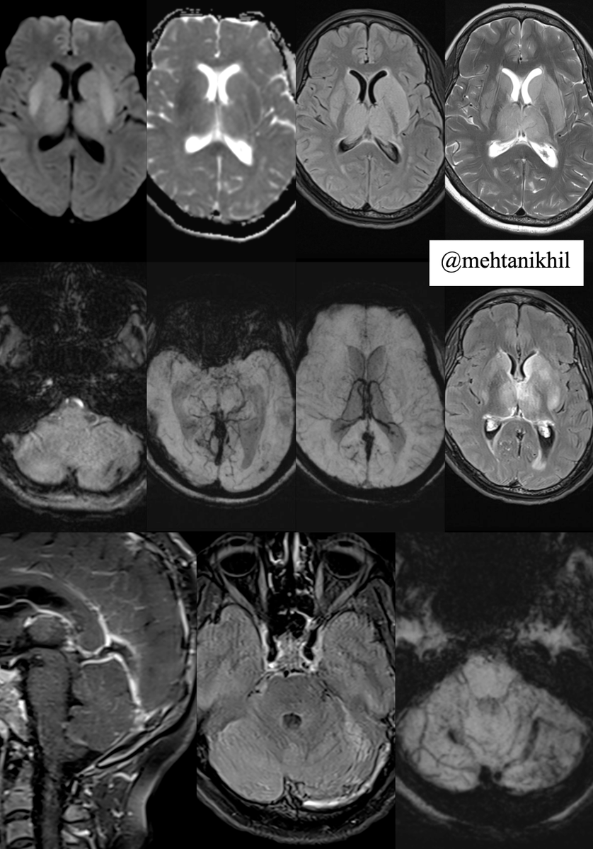
Thank you, sir @nirmalregency. Follow-up CE-MRV: ICV/straight sinus filling defect + CE-FLAIR thrombus in the left transverse sinus. SWI showed cerebellar hemorrhagic venous changes (also in the previous attached images). Pattern fits deep venous thrombosis. Clinical feedback: improving after heparinisation.
Dr. Nikhil R M (NeuroRad) tweet media
English
2
0
1
154
Dr. Nikhil R M (NeuroRad)
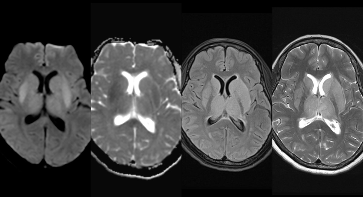
1/2 Case story 🧠 Headache, vomiting, confusion + fever. CT head normal. MRI showed bilateral thalamic–basal ganglia signal abnormality with hippocampal involvement/enhancement. Trap: fever + deep gray lesions can mimic viral encephalitis.
Dr. Nikhil R M (NeuroRad) tweet media
English
4
9
48
6.4K
Dr. Nikhil R M (NeuroRad)
Your question also pushed a useful anatomy point: why relative capsular sparing? Thalami/BG drain dominantly to deep venous channels-ICV/straight sinus. Internal capsule has mixed striate/basal venous drainage + collaterals, so PLIC can be relatively spared while ALIC/genu are involved.
Dr. Nikhil R M (NeuroRad) tweet media
English
1
0
1
69
Dr. Nikhil R M (NeuroRad)
CT scan was done somewhere else, we could only get the report and not the images. Patient came middle of the night for MR Brain and contrast as needed.
English
0
0
0
440
Dr. Nikhil R M (NeuroRad)
Dr. Nikhil R M (NeuroRad)@mehtanikhil·
@nirmalregency Fascinating history! It’s incredible to reflect on how far we’ve come since Alzheimer’s meticulous neuropathologic observations in 1907. Today, we can visualize very plaques & atrophy described with advanced MRI and PET imaging, bridging his foundational work with new diagnostics
English
0
0
1
30
Nirmal Pandey MD DM Neurologist
Nirmal Pandey MD DM Neurologist@nirmalregency·
❸ 1907: Bavarian psychiatrist, Alois Alzheimer is credited w/ the 1st published case of “presenile dementia” in a 51♀ 🔜 his superior Emil Kraepelin termed it as Alzheimer’s disease.
Nirmal Pandey MD DM Neurologist tweet media
English
2
0
3
415
Jack Garnham
Jack Garnham@jjgarnham·
This patient presented with left-sided internuclear ophthalmoplegia (INO). How can we explain this? 👁️👁️ How normal horizontal eye movements work: ⭐️ To look to the right, the brain (mainly from the left frontal eye field) activates the right horizontal gaze center - in the paramedian pontine reticular formation (PPRF) - located in the pons. ⭐️ The right PPRF stimulates the adjacent right abducens nucleus (CN VI). ⭐️ The right abducens nucleus does two things simultaneously: 1⃣ It sends signals to the right lateral rectus muscle → right eye abducts. 2⃣ It sends interneurons that cross the midline and ascend in the left medial longitudinal fasciculus (MLF) to reach the left oculomotor nucleus (CN III, medial rectus subnucleus) in the midbrain. ⭐️ This activates the left medial rectus muscle → left eye adducts. ⭐️ This ensures both eyes move together smoothly to the right! 👁️❌👁️ What happens with a left MLF lesion? ⭐️ A lesion in the left MLF (whether in the pons or midbrain) interrupts the pathway. ⭐️When looking to the right, the right eye abducts normally (intact right CN VI), BUT... ❌The signal fails to reach the left CN III medial rectus subnucleus through the damaged left MLF. ❌Result: impaired adduction of the left eye (it lags behind or stays near the midline). The left MLF is taken out by this infarct!
Jack Garnham tweet mediaJack Garnham tweet media
English
4
20
88
3.4K
Dr. Nikhil R M (NeuroRad)
Dr. Nikhil R M (NeuroRad)@mehtanikhil·
@nodisability @nirmalregency We keep DWI / GRE / Zero TE and Dixon in the optional list of the spine protocol with a short pause after the routine scan protocol completes; which is added depending on the clinical details or imaging findings.
English
1
0
0
28
Nasa
Nasa@nodisability·
@nirmalregency Are we regularly ask for DWI/ ADC for spinal cord? I insist in suspected vascular etiology
Egmore Nungambakkam, India 🇮🇳 English
1
0
1
34
Nirmal Pandey MD DM Neurologist
Nirmal Pandey MD DM Neurologist@nirmalregency·
Hyperacute flaccid typically a bit asymmetric quadriparesis w/ preserved post column: a rarity. Excellent case presentation. Sometimes the cord also needs a DWI screening.
Dr. Nikhil R M (NeuroRad)@mehtanikhil

🧠Case Twist: An adolescent presented with acute flaccid quadriparesis. Initial clinical differential from pediatric neurology: • Viral anterior horn myelitis / AFM • Inflammatory myelitis • Spinal cord infarction #Neuroradiology #PedsNeuro

English
3
1
7
1.8K
Dr. Nikhil R M (NeuroRad)
Dr. Nikhil R M (NeuroRad)@mehtanikhil·
@nirmalregency Yes, DWI of cord was done on the follow1up which lost the restriction window. The prior outside institution scan DWI spine was done, however was ambiguous due to artefacts!
English
0
0
0
82
Dr. Nikhil R M (NeuroRad)
Dr. Nikhil R M (NeuroRad)@mehtanikhil·
Teaching pearl: Anterior horn involvement ≠ always AFM. In acute flaccid paralysis, always integrate: • Clinical clock • Pain at onset • Exertional trigger • DWI/ADC restriction • Vascular topography • Follow-up evolution
English
1
3
3
248
Dr. Nikhil R M (NeuroRad)
Dr. Nikhil R M (NeuroRad)@mehtanikhil·
🧠Case Twist: An adolescent presented with acute flaccid quadriparesis. Initial clinical differential from pediatric neurology: • Viral anterior horn myelitis / AFM • Inflammatory myelitis • Spinal cord infarction #Neuroradiology #PedsNeuro
English
1
4
12
3K