Vicente Martín

3.6K posts

Vicente Martín banner
Vicente Martín

Vicente Martín

@vmargar

NRx.HUNSC #Neurorad

Katılım Haziran 2011
1.6K Takip Edilen3.1K Takipçiler
Vicente Martín retweetledi
Ryo Kurokawa MD PhD #Rdiag
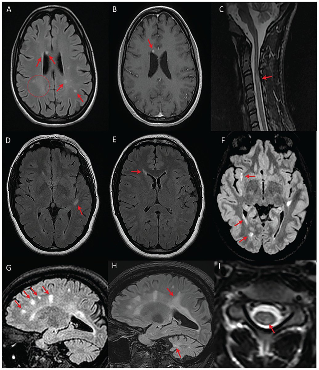
㊗️Adult-type diffuse gliomaの画像診断のまとめ+α、published❗️ ✅秋季大会2025の講演をreview論文化 ✅Astrocytoma vs Oligodendrogliomaの鑑別最新版 ✅Molecular GBMの画像所見 ✅cIMPACT-NOW update 8–11のまとめ PMID:42159911 (JJR) #Rdiag
Ryo Kurokawa MD PhD #Rdiag tweet mediaRyo Kurokawa MD PhD #Rdiag tweet mediaRyo Kurokawa MD PhD #Rdiag tweet mediaRyo Kurokawa MD PhD #Rdiag tweet media
日本語
1
8
48
2.5K
Vicente Martín retweetledi
JAMA
JAMA@JAMA_current·
Amyotrophic lateral sclerosis (#ALS) is an adult-onset neurodegenerative disorder characterized by progressive muscle weakness due to degeneration of upper motor neurons in the brain and lower motor neurons in the brainstem and spinal cord. Diagnosis relies on clinical criteria with mixed upper and lower motor neuron features, and is often supported by electromyography and genetic testing, which may direct treatment. 📌 Learn more in this JAMA Review: ja.ma/3Rc4WVS
JAMA tweet media
English
3
96
288
16.9K
Vicente Martín retweetledi
Maryam Fotouhi, MD
Maryam Fotouhi, MD@Maryamfotouhi11·
#RadInTraining #Tweetorial 1/16 Clinical MRI Biomarkers to Differentiate Parkinson's Disease from Its Mimics 🧠 ✨ Parkinson's disease is still primarily a clinical diagnosis, but MRI can add meaningful diagnostic value when interpreted beyond “no acute intracranial abnormality.”
Maryam Fotouhi, MD tweet media
English
6
65
233
18.8K
Vicente Martín retweetledi
SPIN | Society of Pediatric Neuroimaging
Spin Tweetorial Wednesday Wiskott–Aldrich Syndrome: The great tumor mimic. 📌 Young patient with immunodeficiency + thrombocytopenia 📌 Presents with progressive headache
SPIN | Society of Pediatric Neuroimaging tweet media
English
9
26
115
6.2K
Vicente Martín retweetledi
Dr sthanu subramanian
Dr sthanu subramanian@drsthanus·
Hyperintense vessels seen on a FLAIR MRI scan are a marker of altered blood flow (hemodynamics), not a blood clot (thrombus). *Proximal Indicators: Serpiginous hyperintensities indicate slow forward blood flow or near occlusion of a vessel. *Distal Indicators: These signs suggest retrograde collateral blood flow in the penumbra territory. *Clinical Significance: In the early stroke window (<6 hours), these markers often represent viable penumbra; however, persistent markers may indicate ongoing occlusion and higher risk without reperfusion. @WorldStrokeEd
Dr sthanu subramanian tweet media
English
1
23
97
4.5K
Vicente Martín retweetledi
Lorenzo Pinelli
Lorenzo Pinelli@LorenzoPinelli·
@samrad77 @ongbenghooi1 This is especially true for deep (internal, subcortical) border zone infarcts, which are caused mainly by hemodynamic compromise (whereas superficial/external/cortical border zone infarcts per se are mainly associated to embolic mechanism). 👍🏼 paper -> pubmed.ncbi.nlm.nih.gov/21918038/
Lorenzo Pinelli tweet mediaLorenzo Pinelli tweet mediaLorenzo Pinelli tweet mediaLorenzo Pinelli tweet media
English
0
5
26
976
Vicente Martín retweetledi
Sameer Raniga
Sameer Raniga@samrad77·
When deep (centrum semiovale) + cortical border zone infarcts coexist on the same side, the pattern strongly points to hemodynamic compromise, not embolic shower. Check the ipsilateral vessels. Look carefully at the carotid bifurcation and proximal ICA for high-grade stenosis. In this case, the answer sat at the carotid bifurcation in the neck with high-grade stenosis. Intracranial vessels were clean. Pitfall: Labeling this as “embolic infarcts” and stopping there. You risk missing the real problem. Wisdom: Watershed pattern is a physiology clue. Follow it. — Pearls, pitfalls and wisdom from Today’s reporting list
Sameer Raniga tweet mediaSameer Raniga tweet media
English
4
39
180
8.5K
Vicente Martín retweetledi
Doc Navarrow
Doc Navarrow@DocNavarrow·
💡Lentiform fork sign on MRI typically points to uremic encephalopathy… but not always. ✅Normal renal function should raise suspicion. In this case, CSF revealed anti-NMDA receptor antibodies → autoimmune encephalitis. doi.org/10.1016/j.radc…
Doc Navarrow tweet media
English
1
10
30
1K
Vicente Martín retweetledi
Jack Garnham
Jack Garnham@jjgarnham·
Lovely example of a right-sided Bell's palsy. Intense enhancement of the right fundal, labyrinthine, geniculate, and proximal tympanic segments of the right facial nerve (with the normal left facial nerve for comparison). Remember that, contrary to traditional teaching, you can see mild enhancement of various facial nerve segments in normal patients. Asymmetry and intensity are clues that the enhancement might be pathological.
Jack Garnham tweet mediaJack Garnham tweet media
English
2
18
96
5.5K
Vicente Martín retweetledi
Oliverio
Oliverio@LuisOliverio·
Recién salidas del horno: Guía AHA, 2026, de Evento Vascular Cerebral (EVC) Isquémico. Cambios o puntos relevantes: 🔴 Tenecteplase vs. Alteplase (Cambio Mayor) Se recomienda el uso de Tenecteplase (0.25 mg/kg, bolo único, máx. 25 mg) como una alternativa de primera línea, no inferior a la Alteplase, para pacientes elegibles en la ventana de 4.5 horas. Nota: La dosis de 0.4 mg/kg de Tenecteplase no se recomienda por falta de beneficio adicional y mayor riesgo de seguridad. 🔴 Ictus Leve No Incapacitante: En pacientes con déficits leves que no son incapacitantes (ej. síndrome sensitivo aislado, NIHSS bajo sin impacto funcional) dentro de las 4.5 horas, no se recomienda la trombolisis IV. Se prefiere la doble antiagregación plaquetaria (DAPT). 🔴 Trombectomía (EVT) en "Large Core": Se amplía la indicación de trombectomía mecánica para incluir pacientes con infartos de núcleo isquémico grande establecidos (ASPECTS 3-5), (antes eran excluidos). 🔴 Se desaconseja la reducción intensiva de la PAS (<140 mmHg) tras una reperfusión exitosa. Objetivo: TA <180/105 mmHg en las primeras 24 horas. 🔴 Oclusión de Arteria Basilar: realizar trombectomía en pacientes con oclusión de la arteria basilar a las <24 horas si NIHSS ≥10. 🔴 Control Glucémico: mantener entre 140-180 mg/dL. 🔴 Ventanas Extendidas de Trombolisis: Se recomienda el uso de trombolisis IV en ventana extendida (4.5 a 9 horas o ictus del despertar) en pacientes seleccionados mediante imagen avanzada que demuestre mismatch (DWI-FLAIR o Perfusión). 🔴 Nuevas Guías Pediátricas: Por primera vez se incluyen recomendaciones específicas para pediatría Vamos a revisarlas a fondo para actualizar las apps.
Oliverio tweet mediaOliverio tweet media
Español
7
589
2.1K
84.3K
Vicente Martín retweetledi
Lea Alhilali, MD
Lea Alhilali, MD@teachplaygrub·
4/In the sagittal plane, the nerve looks like an elephant’s trunk coming out of the pons. It should have a smooth curve up and over before it enters Meckel’s cave, just like the way an elephant’s trunk curves.
Lea Alhilali, MD tweet media
English
1
4
12
826
Vicente Martín retweetledi
Neurology Journal
Neurology Journal@GreenJournal·
This Teaching NeuroImage case illustrates an acquired midbrain cleft in a professional boxer, believed to result from repetitive head trauma rather than a single high-intensity traumatic event: hubs.la/Q03-gQVR0 #NeurologyRF @gmneurorx
Neurology Journal tweet media
English
2
21
80
4.7K
Vicente Martín retweetledi
Dr. Brandon Beaber
Dr. Brandon Beaber@Brandon_Beaber·
Are these MRI findings in people with multiple sclerosis? Shockingly, NO! They actually have Wolfram syndrome (WFS), a rare genetic disorder associated with pathogenic variants in WFS1 and is linked to optic atrophy, hearing loss and diabetes. frontiersin.org/journals/neuro…
Dr. Brandon Beaber tweet media
English
9
48
167
22.7K
Vicente Martín retweetledi
Dr. Brandon Beaber
Dr. Brandon Beaber@Brandon_Beaber·
Paramagnetic rim lesions in multiple sclerosis (which appear dark on SWI sequence and are often periventricular) are associated with nearly triple the risk of disability progression (OR 2.87; 95 % CI: 1.01–8.1; p = 0.05) pubmed.ncbi.nlm.nih.gov/41205558/
Dr. Brandon Beaber tweet media
English
0
12
29
2K