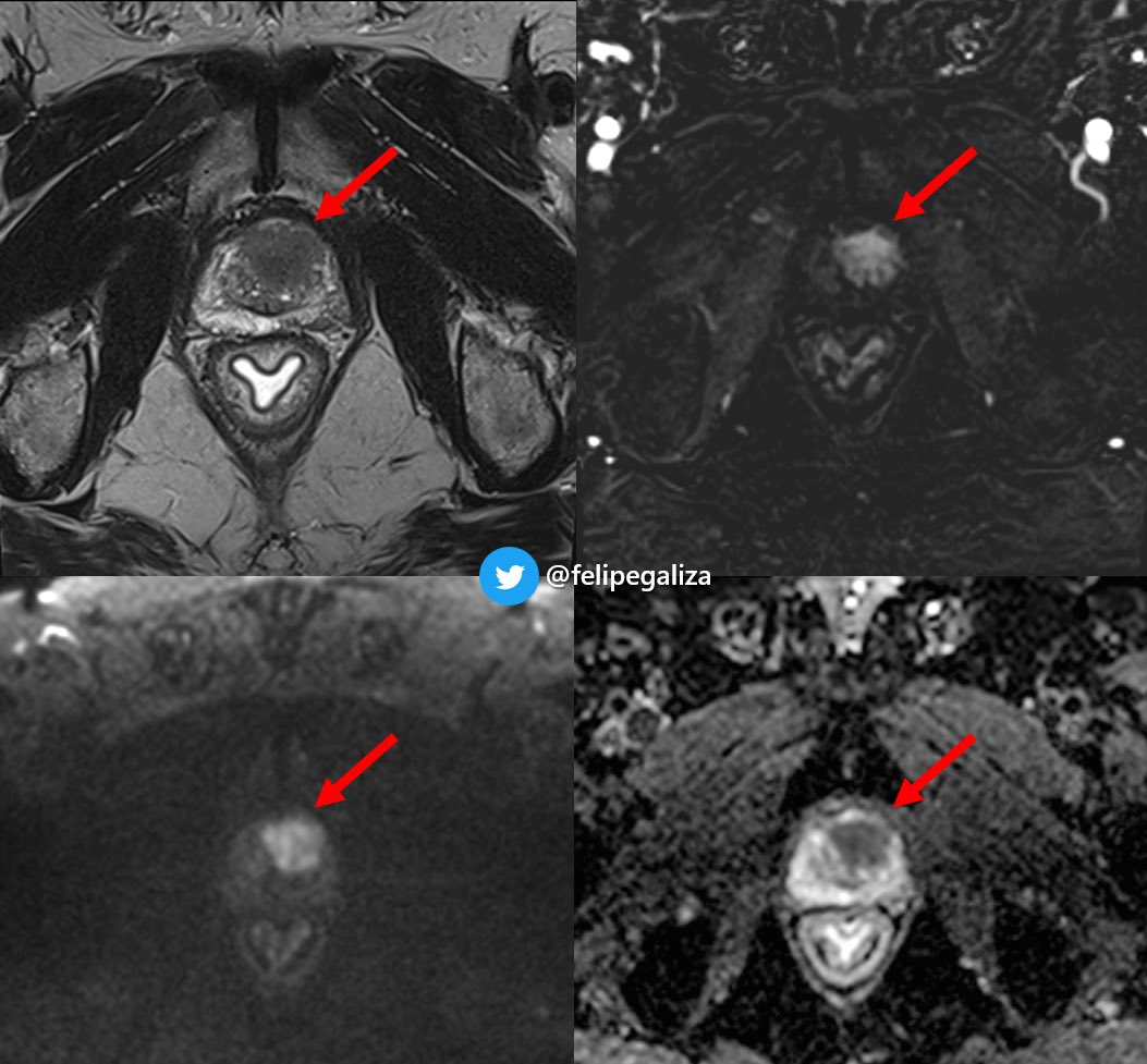
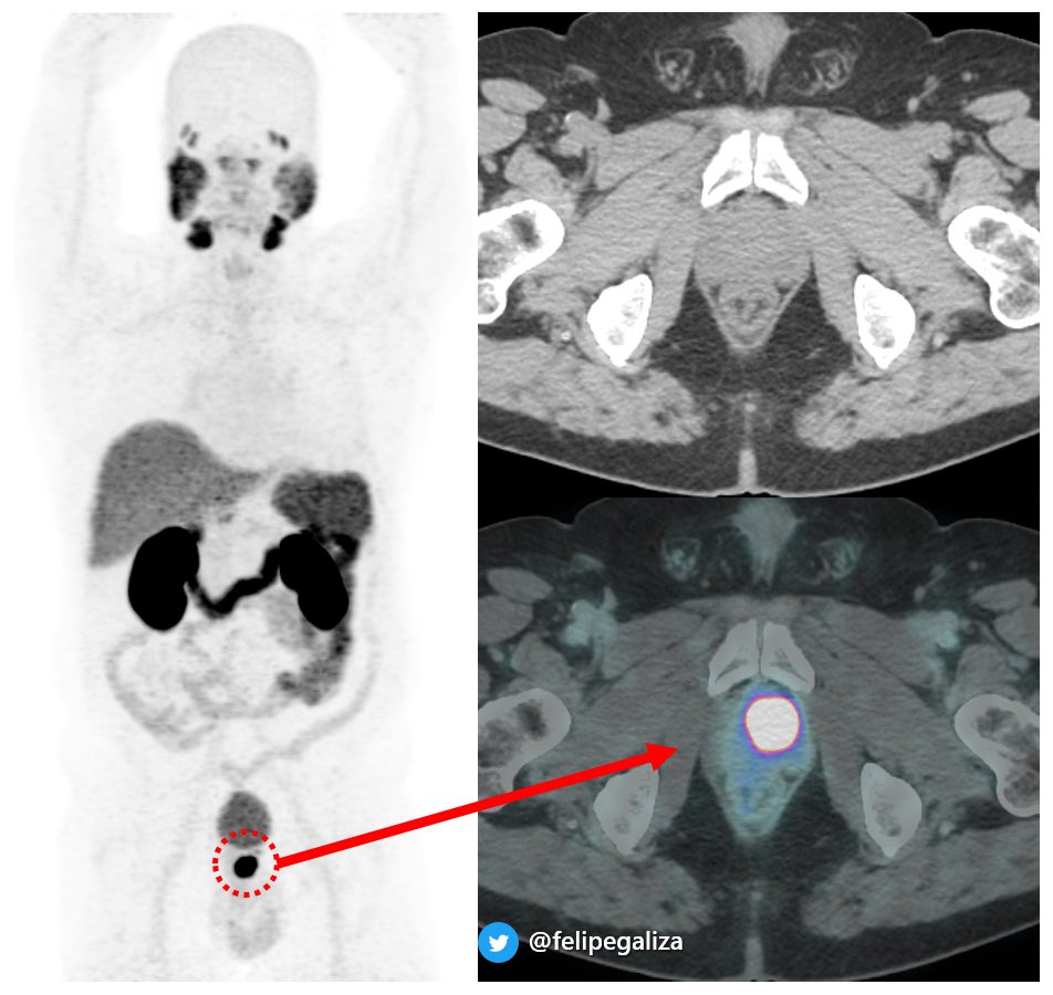
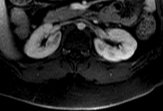
Pinned Tweet
Fernando Ide Yamauchi
245 posts

Fernando Ide Yamauchi
@NandoIde
Abdominal Radiologist, son, husband and dad.
São Paulo, Brasil Joined Ekim 2009
391 Following432 Followers

@danatsouza @LetDdiceFlyHigh @SocietyAbdRad @hiltonmlf @NickSchiedaMD no radiation... i was thinking something like glomerular pathology , but haven't seen any...😂
English

@NandoIde @LetDdiceFlyHigh @SocietyAbdRad @hiltonmlf @NickSchiedaMD Interesting… Any chance she has had radiation to the spine in the last!? That’s the usual explanation for this finding… unlikely in an asymptomatic 14yo…
English

14 y/o female, work up for adrenal adenoma (otherwise asymptomatic).
Has anyone seen this pattern of bilateral renal band of high T2WI, no restriction on ADC, slightly hipoperfused?
@danatsouza @LetDdiceFlyHigh @SocietyAbdRad @hiltonmlf @NickSchiedaMD


English

#AOCR2025 in Chennai was an incredible event!
I had the opportunity to represent CBR at the International Forum on Green Radiology, speak at the BRICS session on bladder cancer staging, and had the honor to serve as Quiz Master for the abdominal imaging session of the International Residents Competition Finals, with teams from over 20 countries.
Grateful for the experience and global friendships during my short and intense passage through India 🇮🇳!
Congratulations and thank you to the organizers from the AOSR and IRIA for the invitation and hospitality.


English

@ErickMRemer @SocietyAbdRad @adrenaldfp There are typical adenomas (homogeneous and <10 UH, probably still safe <20UH).
There are clear "take me out" lesions, large and heterogeneous.
And there are "i don't know" lesions, which unfortunately, there's little to improve with other imaging (CT washout / MRI / PET).
English

42 yo, LUTS, no PSA available (and unfortunately no answer yet...)
Any thoughts?
@SAR_ProstateDFP @Purysko @hiltonmlf @lkayat @danatsouza @CasesCookyJar @radiolobt


English

Recently published paper on postoperative imagining of endometriosis by Neme et.al.
and commented by
@WendalineVB

English

Congresso Imagine 2024 was amazing!
There were many excellent discussions about prostate MRI in several scenarios of prostate cancer.
I want to thank the organizing committee again for the invitation to coordinate the GU schedule alongside my colleague @PublioViana .


Sao Paulo, Brazil 🇧🇷 English

@mauzappa @MorbeckFernando @SchuchAlice @lkayat @IvanPedrollo @Felipegaliza @marcelogusmao2 @CassiaTamura @ivpedrosa @NickSchiedaMD nice case @mauzappa ! as we would say in portuguese "without rodeo": inflammatory lesion. 😂
Now you can hit me with the right answer
English

@coronilRMD @SocietyAbdRad @Felipegaliza @LetDdiceFlyHigh @voxelmator i'll follow @voxelmator , thick walls remind chronic infection. isn't that an excluded upper unit? do we have more coronals?
English

54yo patient, with left renal finding. What’s your opinion? @SocietyAbdRad @Felipegaliza @LetDdiceFlyHigh @voxelmator


English

@Felipegaliza not sure we are ready, but definitely the way to think of a P5: am I confident enough to skip biopsy? If the answer is yes, that's a P5, otherwise, is P4.
English

@Felipegaliza @hiltonmlf @PublioViana @LetDdiceFlyHigh @VChernyakMD @mharisinghani @coronilRMD @SamGalgano @cookyscan1 i'll follow @ankushjaj: saponification of peripancreatic fat
English

@coronilRMD @lkayat @wroclawski_uro we are at the beginning of this journey, learning from @SchuchAlice , @lkayat and many others.
Urethra is retrograde filled with lidocaine (10-15ml). High resolution T2WI are performed, as well as delayed post contrast sequences. finally, dynamic voiding is acquired
English

@lkayat @wroclawski_uro I’m really curious about the acquisition protocol and how you managed to get such beautiful images. 👏🏼👏🏼
English

Is MRI a game-changer to properly investigate urethral stenosis and plan the appropriate treatment? Or retrograde urethrography is enough?
#UroSoMe


Sao Paulo, Brazil 🇧🇷 English

@swoo_rad @epi_rad @SAR_ProstateDFP @radiolobt @Purysko @midrisdoc every time I try to argue this, i get a disapproval from senior colleagues: "if you want to do different from what is stated on the document, than create your own system".
But think about it: weren't we using bosniak on MRI before the updated version?
English

@epi_rad @NandoIde @SAR_ProstateDFP @radiolobt @Purysko @midrisdoc Agree with @epi_rad, but....I do hear what you're trying to say @NandoIde 👂
English

Different case but makes you think of the same question we asked the other day....@SAR_ProstateDFP @radiolobt @Purysko @midrisdoc
Should T2WI be the dominant sequence in TZ followed by DWI (and not consider DCE) as per current PI-RADS? 🤔
Daniel Souza@danatsouza
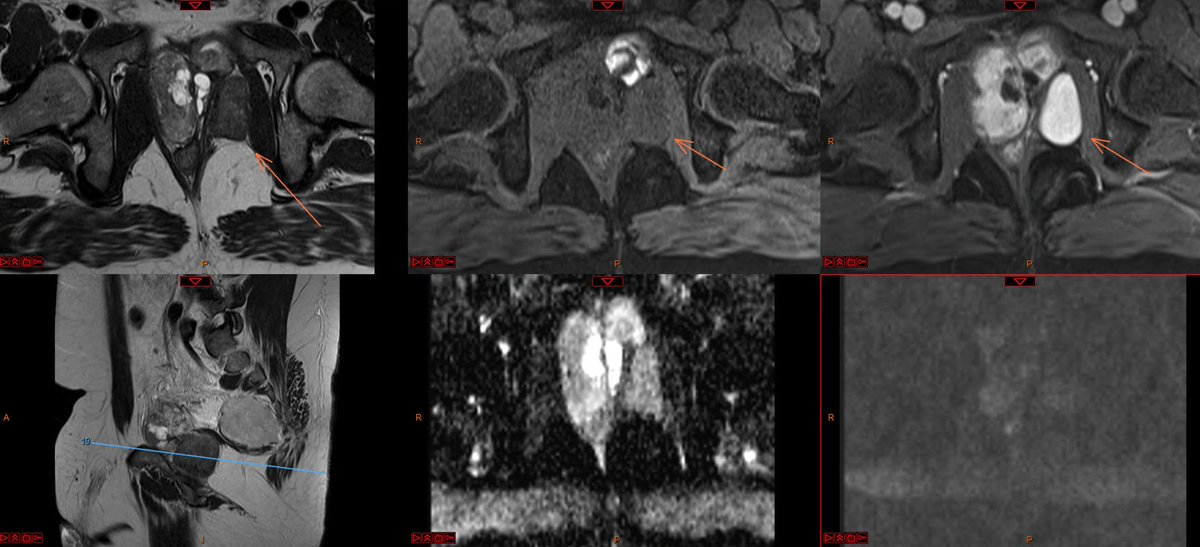
> 1.5 cm T2 dark nodule, right anterior transition zone of the prostate (TZa) + markedly restricting, + DCE by MRI, intense focal uptake at PSMA PET/CT = PI-RADS 5, clinically significant cancer! @AURtweet @FOAMrad @futureradres @CBRadiologia #MedEd @lkayat @SAR_ProstateDFP
English

@Felipegaliza @ErickMRemer @NickSchiedaMD @swoo_rad @hiltonmlf @PublioViana @LetDdiceFlyHigh @coronilRMD i would probably skip formal bosniak, but use concept: no solid component, no thick septa.
calcification = benign
fat = ?
That's a "no touch" lesion, likely to be followed up.
(could that be a calicial diverticulum?)
English

@coronilRMD nice case! chronic hematoma/urinoma? any previous exam to help us?
English

@danatsouza @BrighamRad @AURtweet @FOAMrad @SAR_RFS @futureradres @HarvardMacy @CBRadiologia nice case @danatsouza ! no spleen involvement ?
English

Rare manifestation... what a 'pearl' - pun intended (careful, Dad-iology joke): innumerable punctate calcifications corresponding to peritoneal nodules/granulomas - peritoneal sarcoidosis! @BrighamRad @AURtweet @FOAMrad @SAR_RFS @futureradres @HarvardMacy @CBRadiologia #MedEd

English

@epi_rad there's an old saying that i love to quote: "the best exam is always the prior exam"
English

Just happened to see a similar case here...no history of trauma or pain... 80yo.
Waiting for serum markers, but really favoring a follow up here.

Jonathan A. Hernández Mancera@JHernandez_RX
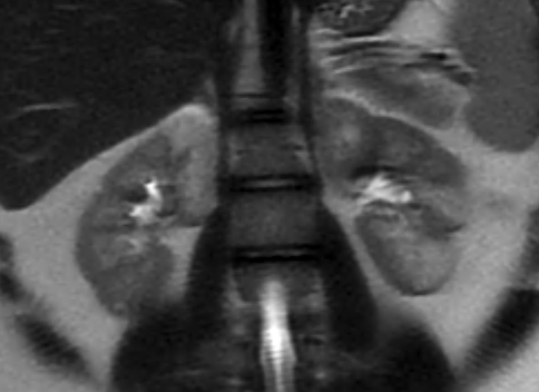
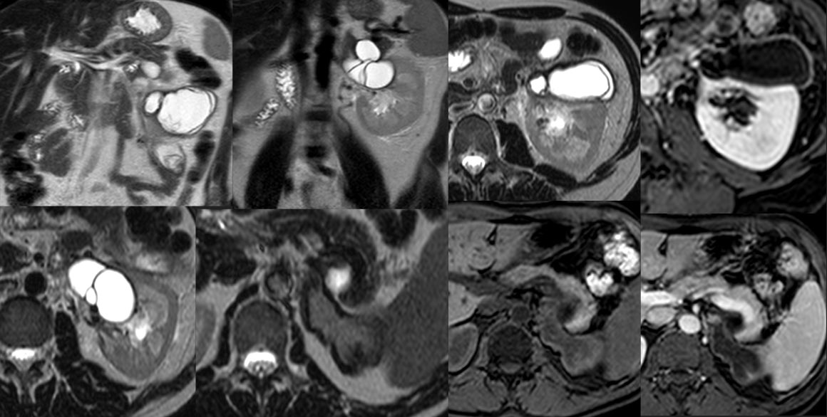
Young patient with right scrotal pain w/o trauma. US shows anechogenic lesion with no Doppler. 2 MRIs 2 months apart show the evolution of a testicular segmentary infarction to a scar. Knowing this pathology could avoid an unnecessary orchiectomy. #radiology #urology
English


