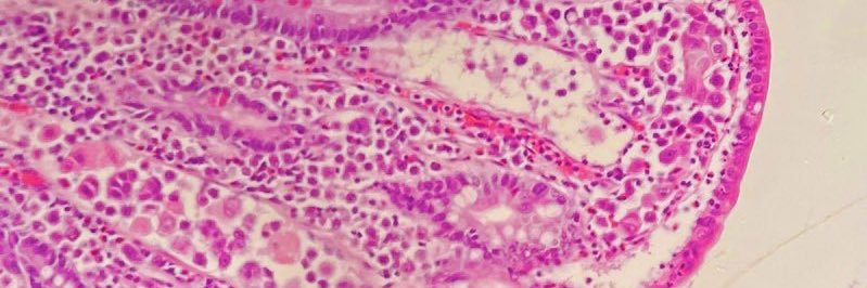
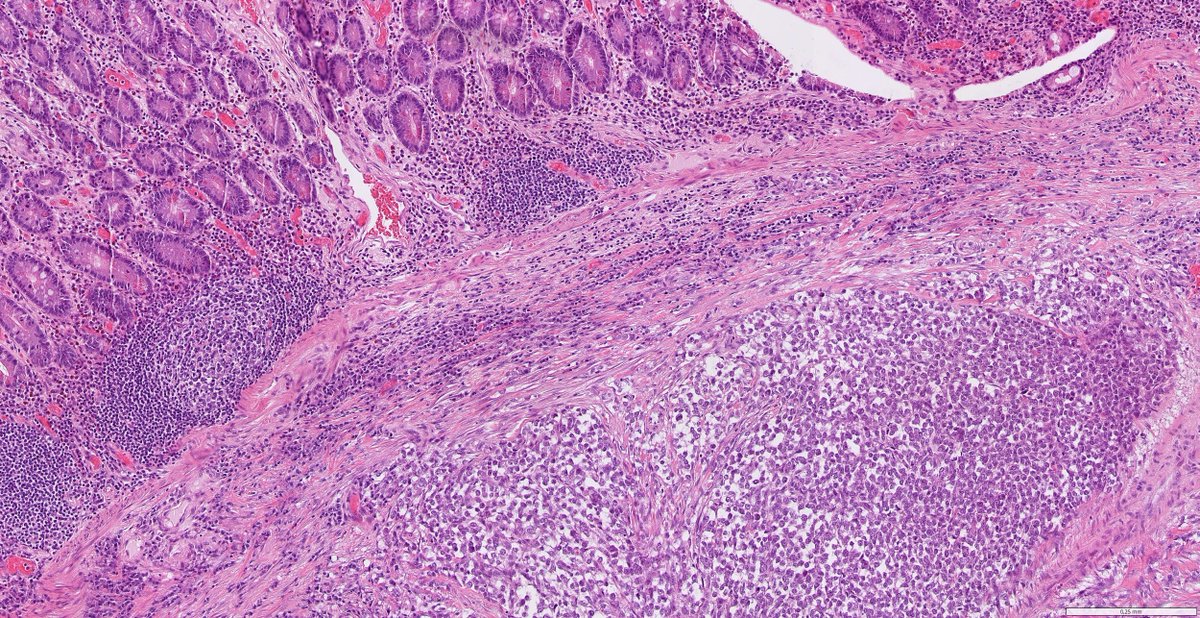
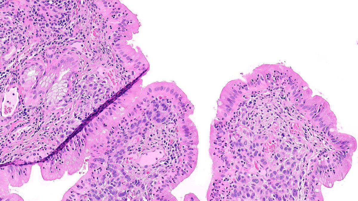
Amal Asar, FRCPath, MAcadMED

3.1K posts

Amal Asar, FRCPath, MAcadMED banner
Amal Asar, FRCPath, MAcadMED

Amal Asar, FRCPath, MAcadMED

@amalasar86

Histopathologist (I see the diseased tissue rather than the diseased human 🙂),Breast and GI pathology. Mom 👦👦 and passionate student . #Pathtweetaward judge.

Katılım Eylül 2009
548 Takip Edilen1K Takipçiler
Amal Asar, FRCPath, MAcadMED retweetledi
Dr. Luca
Dr. Luca@atman_ci·
One of the major educational initiatives in #Pathology, one of the most impactful step for democratization and digitalization of education in Pathology Thanks to you I have learned and continue to learn so much. Heartfelt congrats Dear Friend @mannanrifat03 for the 300th PathCast
Dr. Luca tweet media
English
1
4
29
2K
Amal Asar, FRCPath, MAcadMED retweetledi
C. Christofer Juhlin
C. Christofer Juhlin@DrJuhlin·
Not everything is what it seems. Middle-aged patient. Consult case. Minimal clinical data. Ileal resection for obstruction → tumor found. Let’s follow the diagnostic trail. Short thread, do the polls in order (and no sneak peek...)
C. Christofer Juhlin tweet media
English
2
43
97
6.4K
Olaleke Folaranmi
Olaleke Folaranmi@DrGeeONE·
Power list 2025 I was featured on “The Pathologist” Power List 2025: Leading Voices Edition. It is a privilege to be counted among those considered as “inspirational figures behind innovation and achievement in pathology and laboratory medicine” It didn’t stop there; I was voted one of the readers’ favourites, and I am grateful to you all! My interview has just been published: thepathologist.com/issues/2026/ar… youtu.be/q_svuJTWi28?si… @Nigerian_Doctor @DrJoeAbah @enodamade @DrKamleshDarji1 @Just_JONIER @chriszioga @SumantaDas_7 @pepeheffernan @Baskotacytopath @VarshaManuchaMD @mdlozanoe @01sth02 @LJTafeMD @Dr_Jukic @kriyer68 @LaraHarikMD @NebbacheHafsa @HENRYY_MD @SGottesmanMD @RunjanChetty @HermelinMD @darcykerrMD @sakhurram @Pathmath1 @kis_lorand @edusqo @ariella8
YouTube video
YouTube
Olaleke Folaranmi tweet mediaOlaleke Folaranmi tweet media
The Pathologist@pathologistmag

🚨 The Pathologist Power List 2025 is now LIVE! Meet 50 Leading Voices moving pathology forward with big ideas and bold action. 👏 Celebrate this year’s winners and read their essays: ow.ly/UBJs50X0hzV #ThePathologist #PowerList2025 #Pathology #PathX #PathTwitter

English
33
28
119
17K
Amal Asar, FRCPath, MAcadMED
In this talk at will try to explore how large scale implementation can shift how we work and train as Pathologists, and shed a light on requirements to be respected while working with AI in the hybrid workplace, to maintain patient safety and preserve Pathologists’ identity.
Amal Asar, FRCPath, MAcadMED tweet media
English
0
0
0
58
Amal Asar, FRCPath, MAcadMED retweetledi
Raza Hoda MD
Raza Hoda MD@RazaHoda·
Case of Metastatic Lobular Carcinoma to Gallbladder ☄️🔬 Chronic cholecystitis on the requisition? 🤔 Those dyscohesive cells in the wall might tell a different story 😦 #PathX #PathTwitter #breastpath
Raza Hoda MD tweet media
English
3
18
74
2.7K
Amal Asar, FRCPath, MAcadMED retweetledi
Tim Bracey
Tim Bracey@drtimbracey·
gastric biopsy from an old man what do you think is going on here? #GIpath #PathTwitter
Tim Bracey tweet mediaTim Bracey tweet mediaTim Bracey tweet mediaTim Bracey tweet media
English
9
44
112
10.2K
Amal Asar, FRCPath, MAcadMED retweetledi
Raza Hoda MD
Raza Hoda MD@RazaHoda·
Another frightful feature 🧛‍♂️ haunting pathologists 😱 this spooky season 🎃 Case of Microglandular Adenosis 👻🔬 Small round glands with bright eosinophilic secretions🩸infiltrating fat without myoepithelial cells that mimics invasion. 🧟‍♂️ #PathX #PathTwitter #breastpath
Raza Hoda MD tweet media
English
6
31
102
4.2K
Amal Asar, FRCPath, MAcadMED retweetledi
Aysha Mubeen
Aysha Mubeen@AMubeen_Path·
Not my typical #gupath #gynpath post but an interesting one for trainees! Colon biopsy for GI bleed, clinical concern for IBD. #pathboards
Aysha Mubeen tweet mediaAysha Mubeen tweet media
English
13
62
258
19.7K
Amal Asar, FRCPath, MAcadMED retweetledi
Fouad Boulos
Fouad Boulos@fouad_boulos·
Happy Saturday everyone! Let's discuss our Breast Consult Case of the Week 3 #BCCW #breastpath #PathTwitter Like everyone astutely pointed out, the diagnosis is adenomyoepithelioma (AME). Adenomyoepitheliomas are biphasic tumors of the breast, and they may encompass a variety of patterns, potentially making their diagnosis challenging. In my experience, the most common pattern of adenomyoepithelioma is papillary with a neoplastic population of spindled myoepithelial cells. This is however not the only way these tumors can show up in your practice. As a matter of fact, when the lesion is papillary in nature, one may question whether one is dealing with adenomyoepithelioma or an intraductal papilloma with myoepithelial hyperplasia. I believe the key to diagnosing these lesions is to recognize the neoplastic nature of the myoepithelial cells, both morphologically and quantitatively, meaning that they should be increased in number independently of the epithelium, and the myoepithelial cells themselves should be morphologically abnormal (enlarged, potentially with prominent nucleoli, often with a clear cytoplasm). In the above case, the myoepithelial cells just appear enlarged and spindled, and growing in what appears to be a clonal process. One of the main controversies linked to adenomyoepitheliomas is their reported potential to behave in a malignant fashion without necessarily showing malignant histology. This is fortunately exceedingly rare. What I often observe however is the potential progression of adenomyoepithelioma to atypical and malignant forms. Here are some morphologic and immunohistochemical clues to the diagnosis. 1. AME can sometimes consist of infiltrative looking tubules and when you stain it for myoep markers it will be variably positive and hence confusing, so if you don’t think about it you might easily miss it 2. Squamous and sebaceous differentiation are sometimes seen and are a clue to the diagnosis 3. Avoid calling malignant AME if it’s cytologically atypical but well contained. Atypical AME works most of the time 4. Densely hyalinized stroma is common due to deposition by the myoepithelial cells, and often surrounds the whole lesion. Trickling of neoplastic cells beyond the hyalinized stroma might be reason for concern 5. Aberrant staining patterns for myoep markers are common, like in this case where p63 is lost in much of the spindle cells. That’s actually a clue to the diagnosis. The topic is of course highly complex and diverse for a such a rare lesion. The key, I think, is to remember this entity when dealing with a lesion that doesn’t seem to make sense and consider it in your differential diagnosis. Here’s a very good paper by Dr Rakha addressing the many complexities of AME. Rakha E, Tan PH, Ellis I, Quinn C. Adenomyoepithelioma of the breast: a proposal for classification. Histopathology. 2021 Oct;79(4):465-479. doi: 10.1111/his.14380. Epub 2021 Jun 13. PMID: 33829532. @washupathedu @washu_pathology
Fouad Boulos tweet mediaFouad Boulos tweet media
English
1
17
57
3.6K
Rahul Deb
Rahul Deb@RahulADeb·
Sad to hear Dr. Borges is no more. An icon in pathology & a teacher who shaped us all. Her "bring any slide" teaching sessions were masterclasses & lesson to "speak the surgeon's language" stays with me. Her legacy lives on in her students for generations. RIP Ma’am. Thank you
Rahul Deb tweet media
English
1
0
2
165
Dr. Luca
Dr. Luca@atman_ci·
For a #Pathology within the reach of all #pathologists in the world, in complexity one must simplify for the patients good Much thanks Dear Colleagues for this paper: “Proposals to make diagnostic criteria truly usable and useful to pathologists worldwide” jcp.bmj.com/content/early/…
English
3
12
26
1.5K
Amal Asar, FRCPath, MAcadMED retweetledi
Mateen Hussain
Mateen Hussain@MateenH79781868·
So I start my morning today with these guys....
Mateen Hussain tweet media
English
3
7
41
4.6K
Amal Asar, FRCPath, MAcadMED
Amal Asar, FRCPath, MAcadMED@amalasar86·
Enjoying the day so far at the brilliant London GIT course where a fantastic line up of local and international experts are sharing wisdom on various treacherous GI topics @London_GI_Path Looking forward to a week of knowledge and fruitful conversations. #pathtwitter #GIPath
Amal Asar, FRCPath, MAcadMED tweet mediaAmal Asar, FRCPath, MAcadMED tweet media
English
2
0
2
152
Amal Asar, FRCPath, MAcadMED retweetledi
Mark Ong
Mark Ong@DrMarkOng·
The different faces of immunoglobulin in B-cell and plasma cell neoplasms. They can deposit as crystals. Tweet below was a DLBCL, this picture was a plasma cell myeloma. twitter.com/DrMarkOng/stat… #hemepath #pathology
Mark Ong tweet media
Mark Ong@DrMarkOng

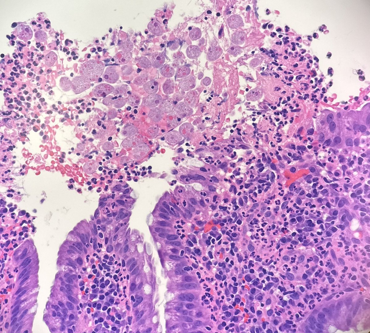
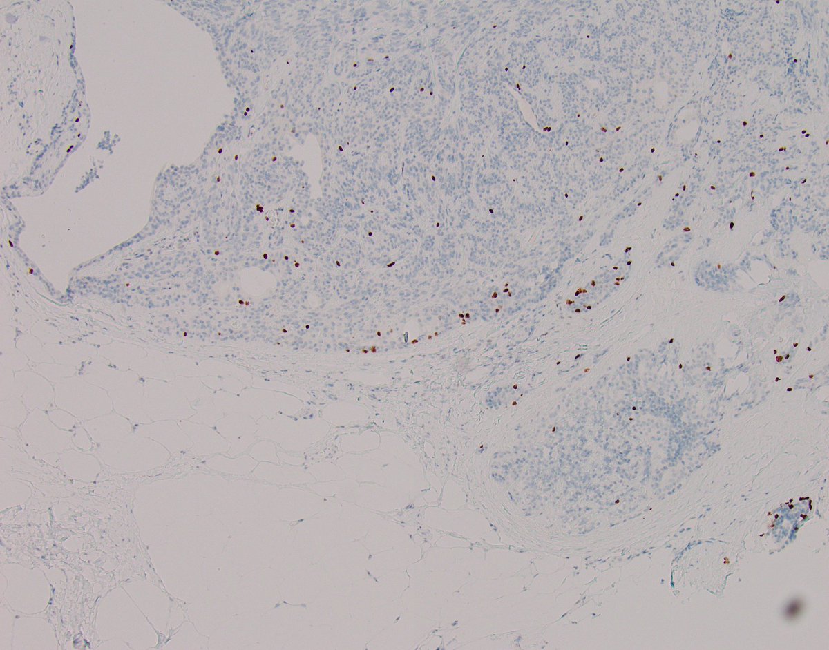
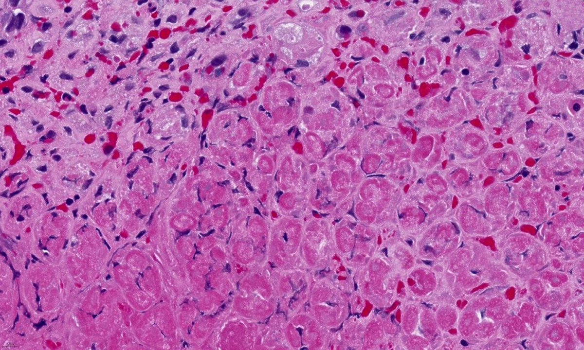
Crystals arising in within a high grade B-cell lymphoma (called DLBCL on account of B-cell markers). If I had this case today, I'd do a range of plasmacytic markers as that might tip it into plasmablastic lymphoma. #hemepath #pathology

English
5
21
70
9.8K