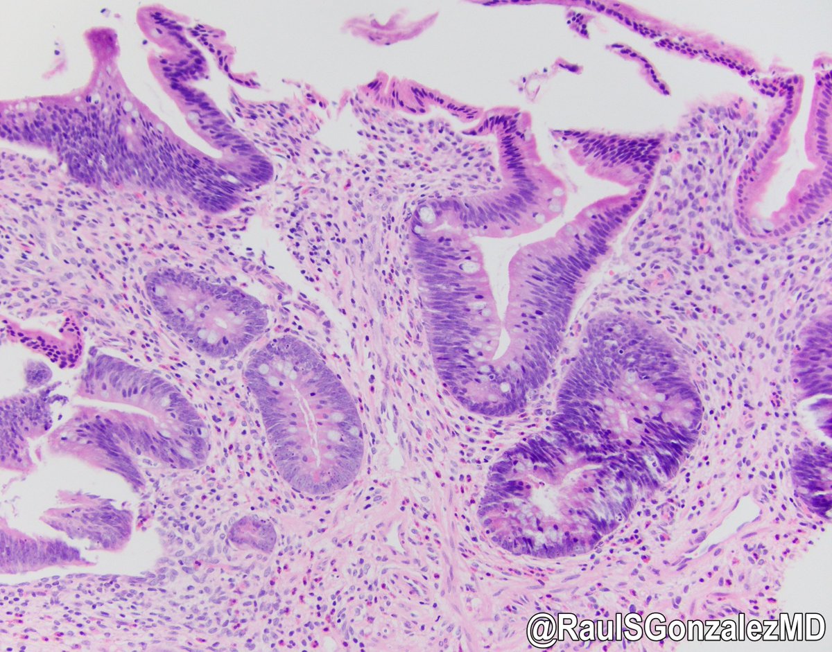
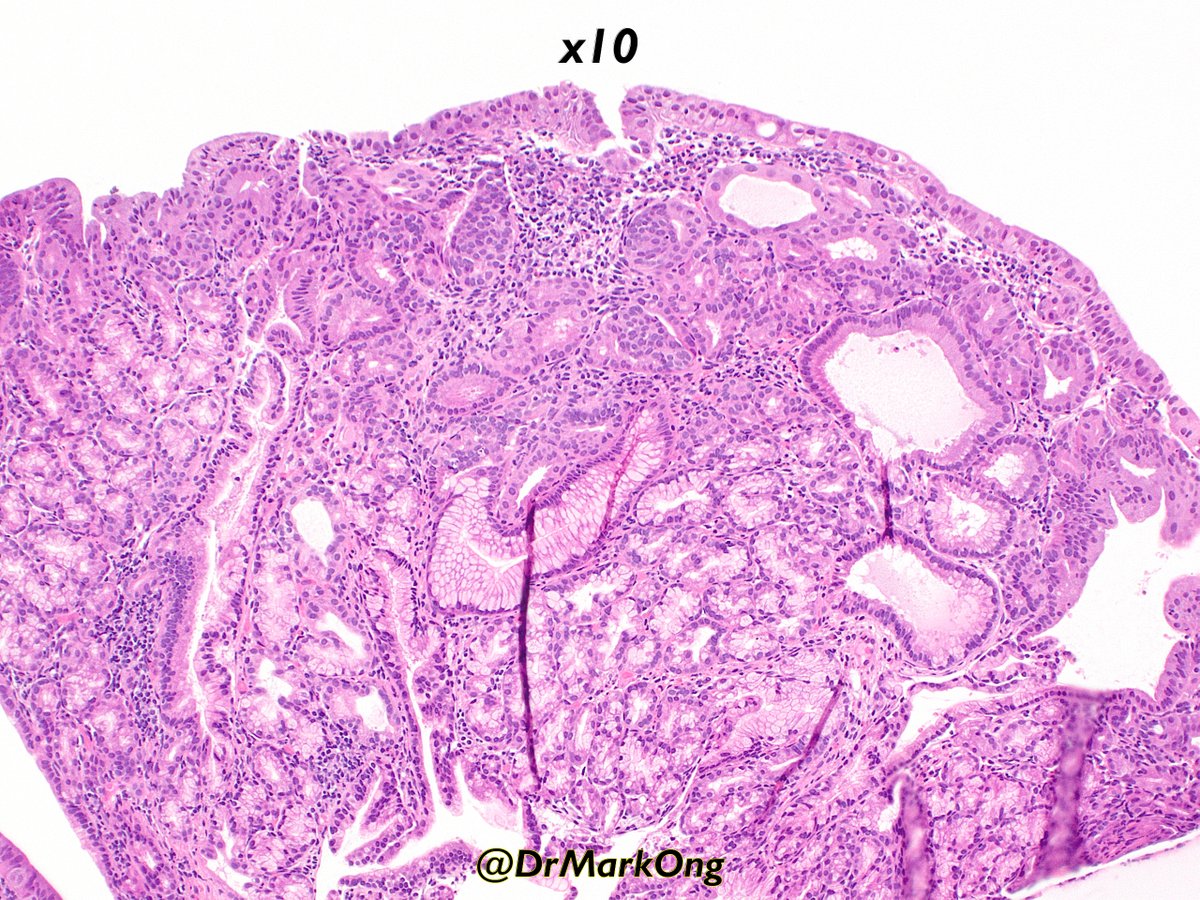
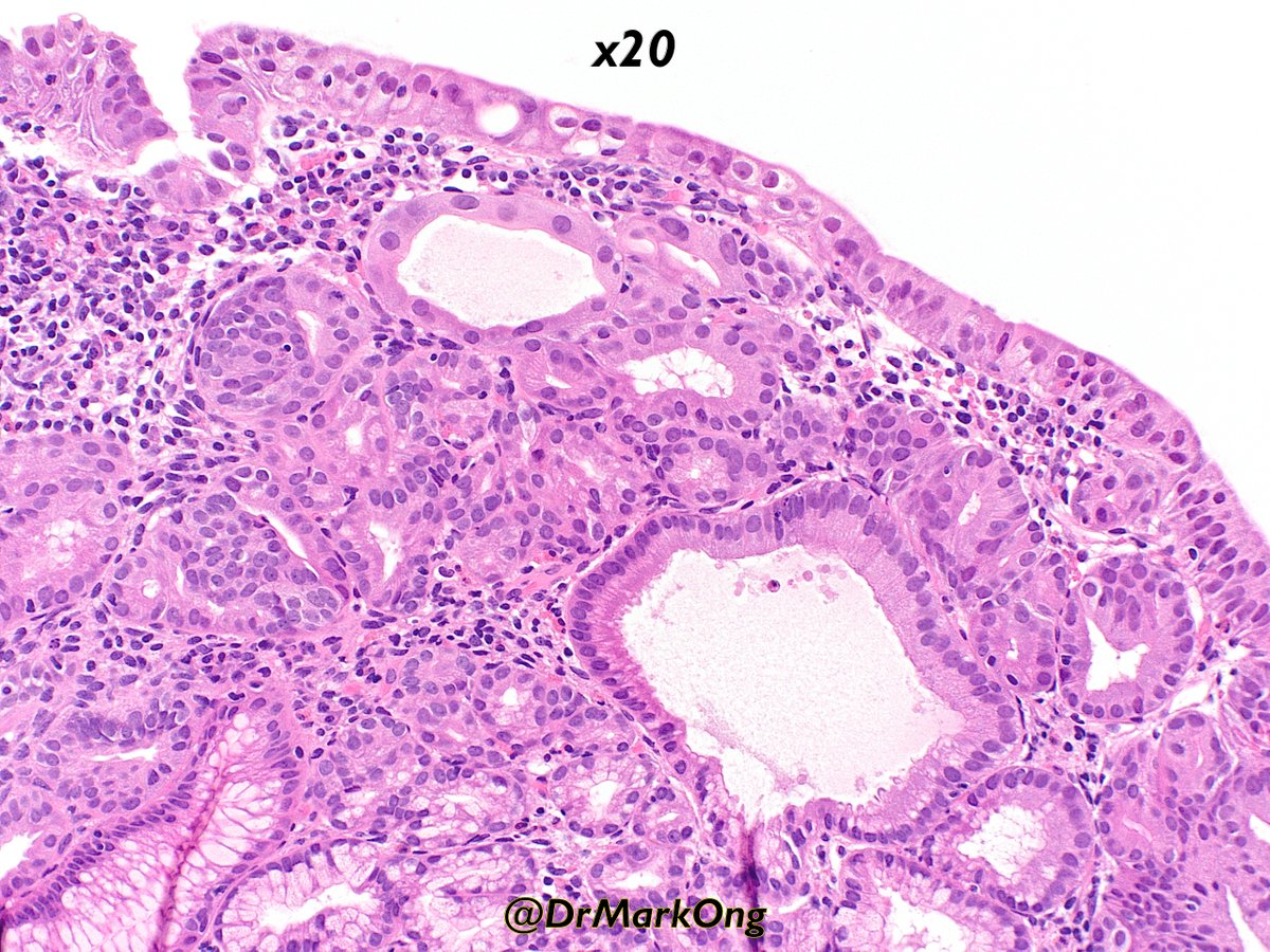
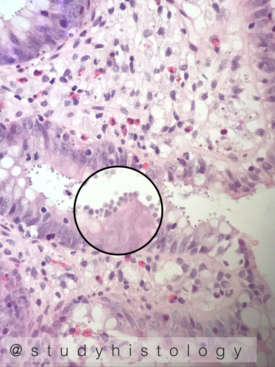
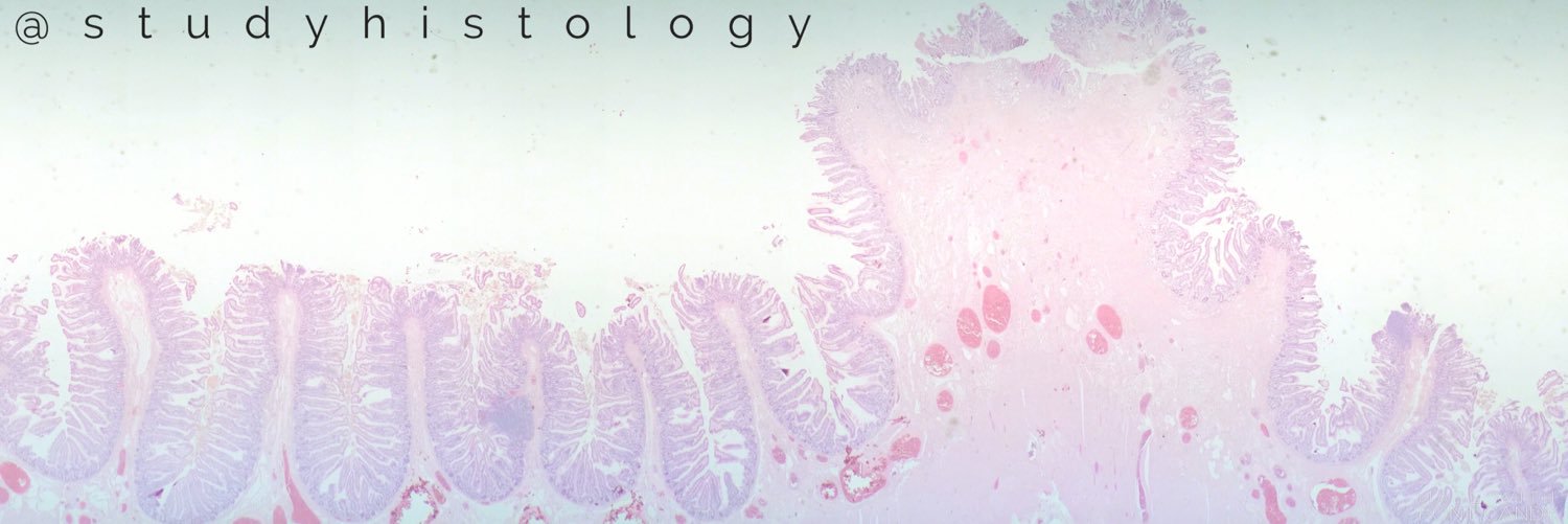
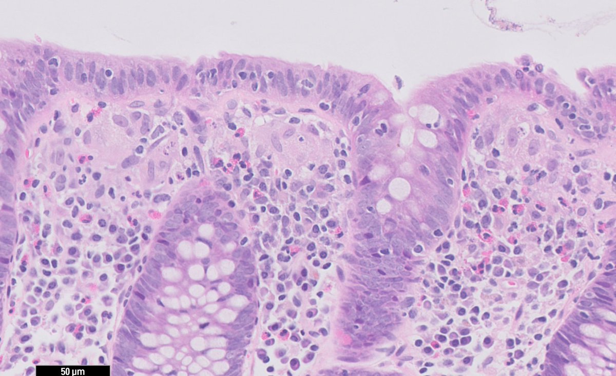
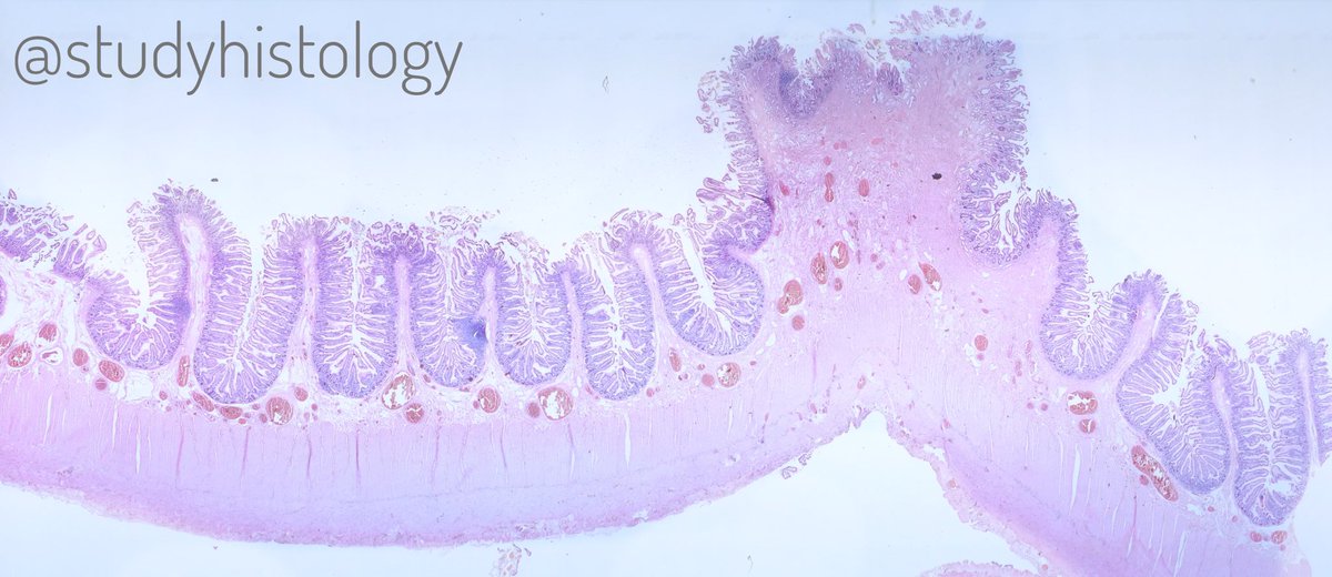
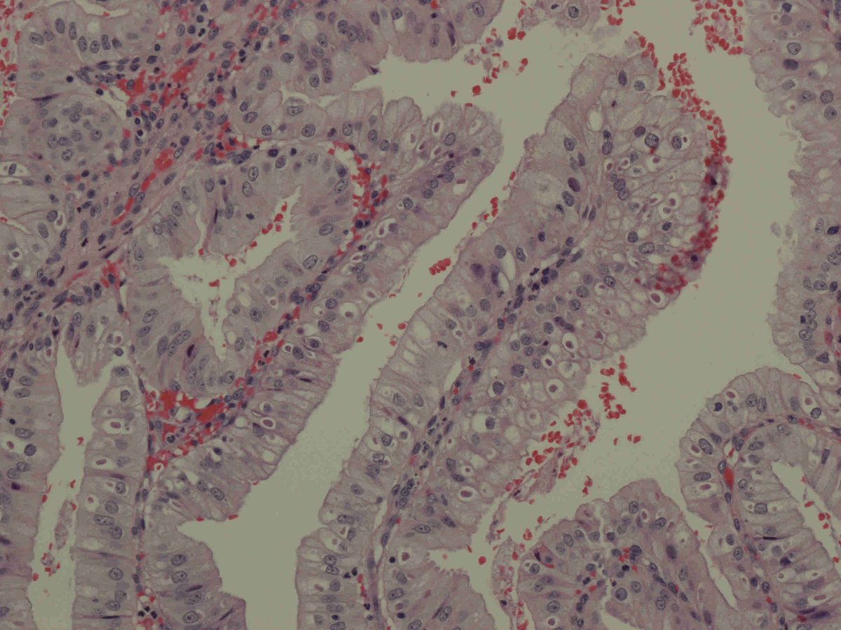
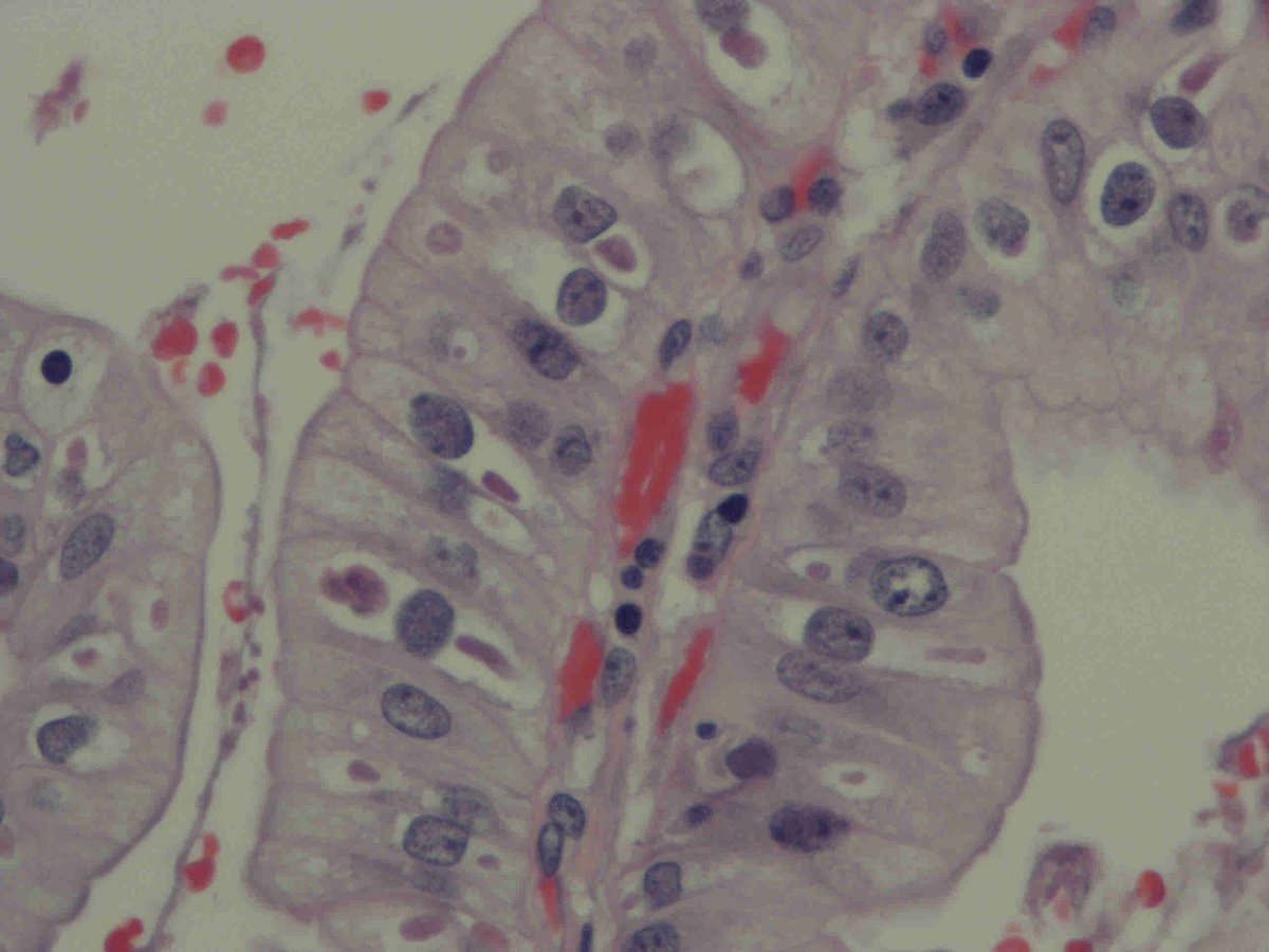
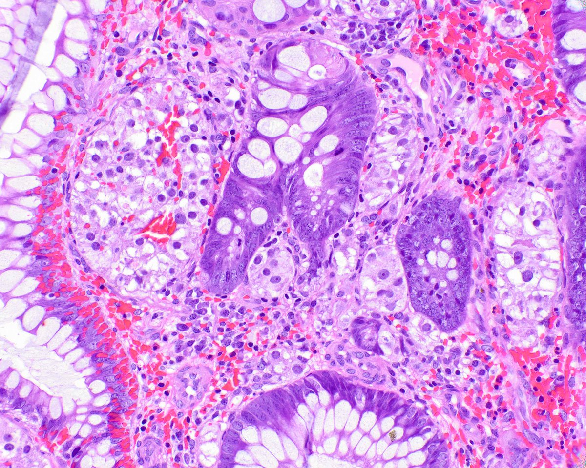
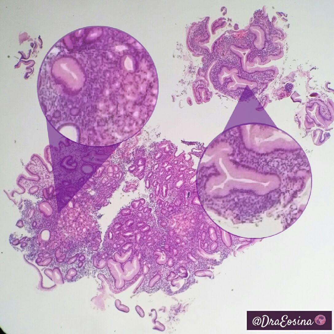
Histology Student

1.6K posts

Histology Student banner
Histology Student

Histology Student

@studyhistology

Passionate about Pathology | #gipath #histopathology🔬| All views are my own | Tweets and Retweets are not Medical Advice
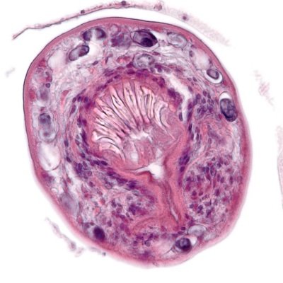
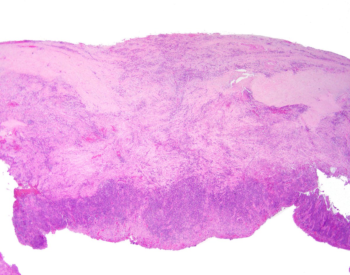
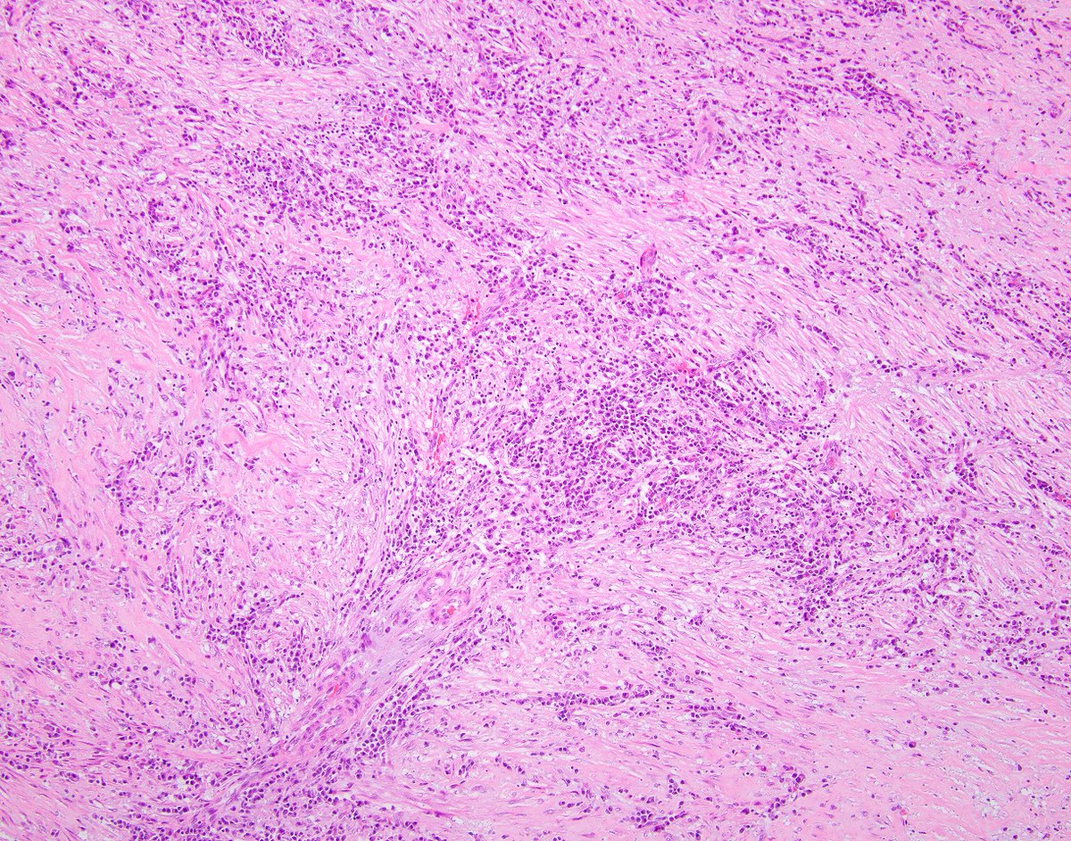
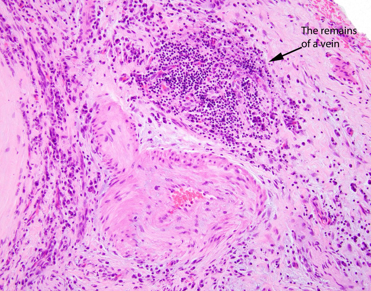
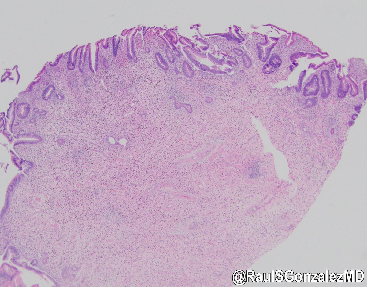
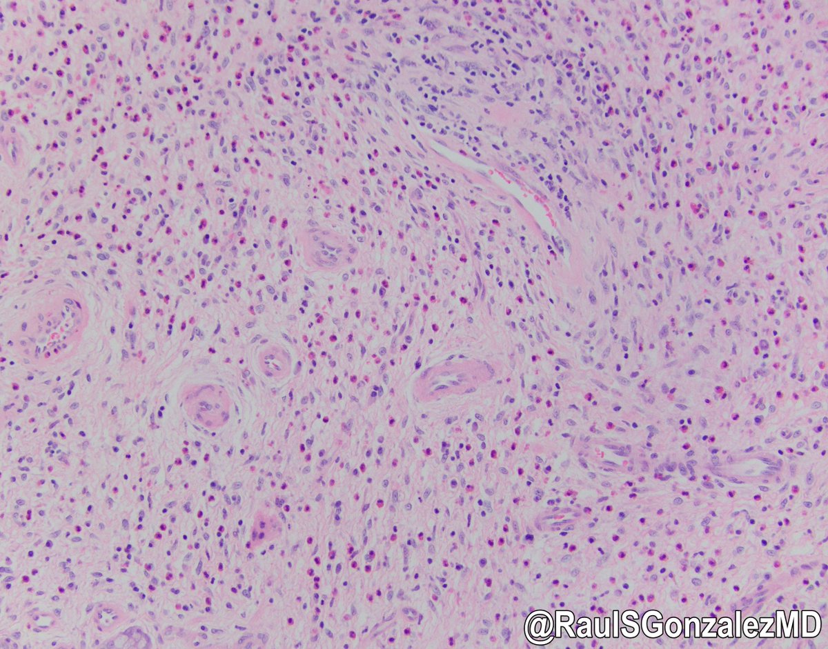
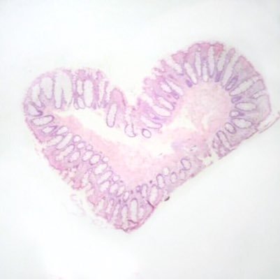
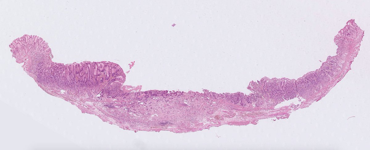
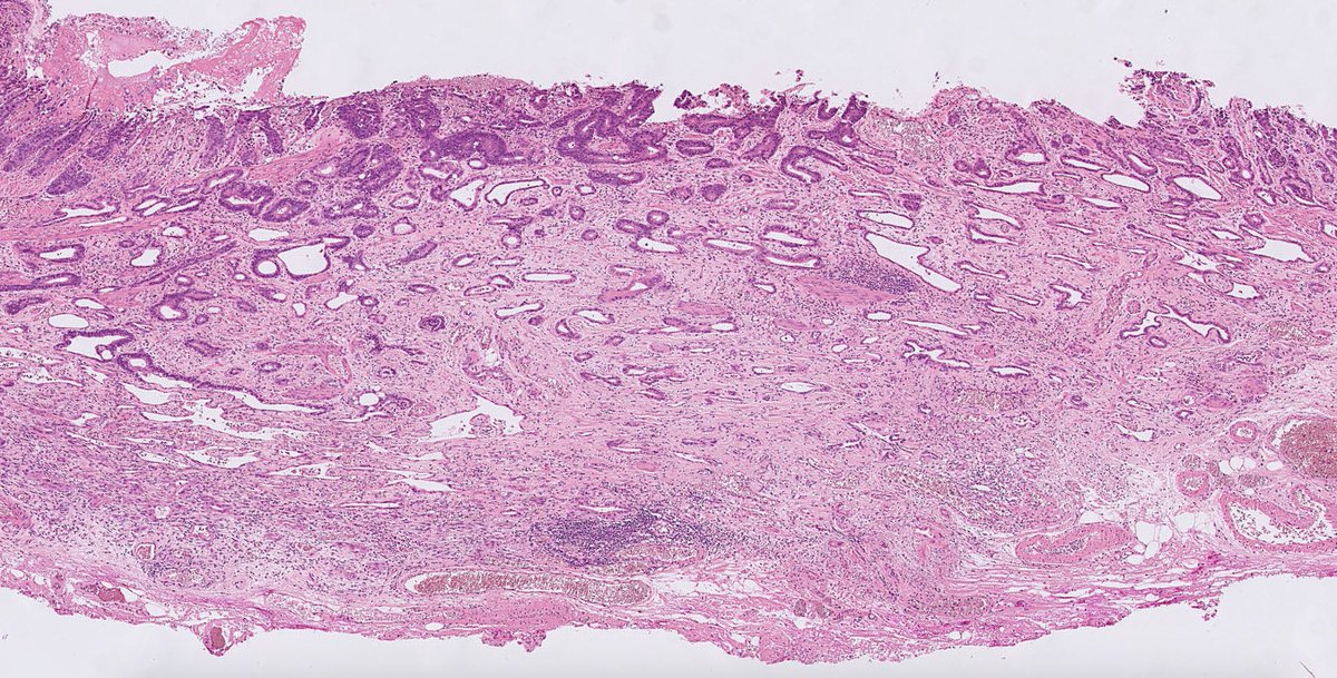
United Kingdom Katılım Ocak 2014
362 Takip Edilen1.9K Takipçiler
Histology Student retweetledi
Marnix Jansen
Marnix Jansen@jansen_marnix·
Here's an odd 🐦 Microscopic colitis pattern, but you find rows of giant cells and macrophages have accumulated in the upper mucosal reaches. Normal colonoscopy, no medication, usual reason to investigate. What's going on? #GIpath
Marnix Jansen tweet mediaMarnix Jansen tweet mediaMarnix Jansen tweet mediaMarnix Jansen tweet media
English
9
50
151
0
Histology Student retweetledi
Mark Ong
Mark Ong@DrMarkOng·
Biopsies of a duodenal D1 polyp. More pictures in next tweet. Would be interested in how people would report this? Perhaps the ultimate label doesn't matter that much as I think it needs removal. #GIpath
Mark Ong tweet mediaMark Ong tweet mediaMark Ong tweet mediaMark Ong tweet media
English
12
26
71
0
Histology Student
Histology Student@studyhistology·
Chronic mucosal injury is often seen overlying the diaphragms including gastric metaplasia (seen here) 2/2
Histology Student tweet media
English
0
0
7
0
Histology Student
Histology Student@studyhistology·
@miss_me1986 Low power view of diaphragm disease highlights the difference in height between the diaphragms and the adjacent normal plicae circularis 🔬
Histology Student tweet media
English
0
0
3
0
Amal Asar, FRCPath, MAcadMED
3/4 * There can be minimal inflammation and architectural distortion in the mucosa overlying or surrounding the diaphragms. The inflammation is usually limited to the submucosa and no granulomas/ lymphocytic aggregates are usually detected; an important point to keep in mind.
Amal Asar, FRCPath, MAcadMED tweet media
English
2
3
29
0
Histology Student retweetledi
Marnix Jansen
Marnix Jansen@jansen_marnix·
Submucosal infiltration in ESD specimens often provokes extensive fraying of the muscularis mucosae. Measure infiltration depth from deepest fibre MM, true volume of disease often larger than raw depth indicates. Here 620 micron, T1b SM2, 15% risk LNs gut.bmj.com/content/68/9/1…
Marnix Jansen tweet mediaMarnix Jansen tweet mediaMarnix Jansen tweet mediaMarnix Jansen tweet media
English
2
16
44
0
Histology Student retweetledi
Xiaoyin "Sara" Jiang, MD (She/Her)
#PathPitfall, Frozens Edition: We often freeze nodules on the liver looking for mets. When we see a proliferation of glands in the liver, a knee-jerk response is: metastatic adenoca! BUT don't forget our friend bile duct hamartoma- common, often incidentally found, and benign!
Xiaoyin "Sara" Jiang, MD (She/Her) tweet media
English
2
30
104
0
Histology Student retweetledi
Elizabeth Montgomery, MD
Elizabeth Montgomery, MD@LizMontgomeryMD·
Incredible example of a TFE3-rearranged PEComa that presented as a colorectal polyp.
Elizabeth Montgomery, MD tweet mediaElizabeth Montgomery, MD tweet mediaElizabeth Montgomery, MD tweet mediaElizabeth Montgomery, MD tweet media
English
13
164
382
0
Histology Student retweetledi
Laura G. Pastrián MD
Laura G. Pastrián MD@DraEosina·
These 'thickened gastric folds' in a 50yo man showed 4 different diagnoses, all of them benign 😀 🕐 Superficial active gastritis (see those neutrophils! HP present, not shown 🕑 Intestinal metaplasia, non dysplastic (probably related to atrophy) (Cont.) #gipath #pathology
Laura G. Pastrián MD tweet mediaLaura G. Pastrián MD tweet media
English
3
34
67
0
Histology Student retweetledi
Judith Ferry
Judith Ferry@judith_ferry·
Hey #Pathologists, what do you think of this enlarged inguinal lymph node from 62 yo male with CLL? Part of node shows SLL/CLL but part is necrotic and edematous. What’s going on?
Judith Ferry tweet mediaJudith Ferry tweet mediaJudith Ferry tweet media
English
7
46
74
0
Histology Student retweetledi
Raul S. Gonzalez, MD
Raul S. Gonzalez, MD@RaulSGonzalezMD·
Jejunum biopsy. Clearly an unhappy piece of mucosa. What do you see? Or rather, what do you NOT see? #pathology #gipath
Raul S. Gonzalez, MD tweet media
English
11
44
94
0
Histology Student retweetledi
Christina Arnold, MD🦋
Christina Arnold, MD🦋@CArnoldMD·
1 of 2 1D Diagnosis: Esoph leukoplakia/epidermoid metaplasia Pearls: -endoscopic findings: plaque (may come as "r/o candida"); if diffuse, consider Tylosis -histologic criteria: orthokeratosis & granular layer
Christina Arnold, MD🦋 tweet media
Christina Arnold, MD🦋@CArnoldMD

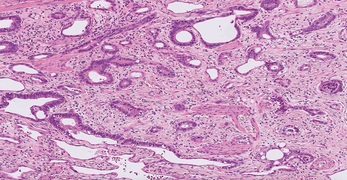
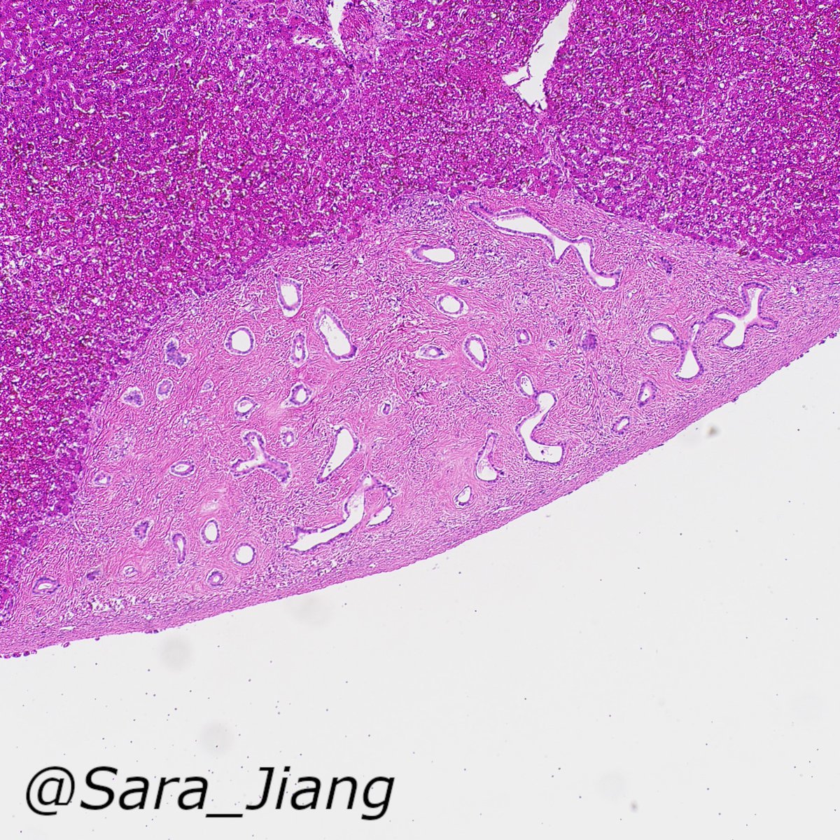
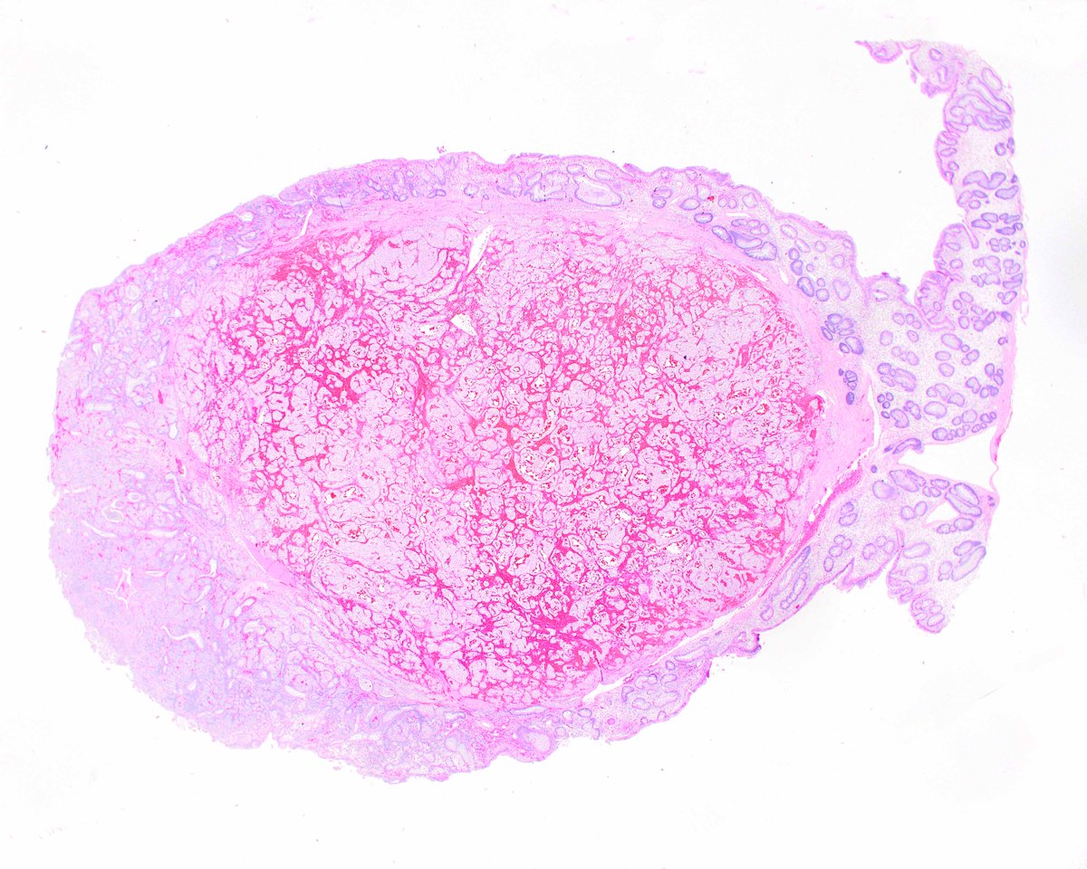
Bravo #RedHotChiliPeppers! Round 1 answers: 1D, 2A, 3C, 4E Round 2 challenge: Submit the diagnoses for the 4 cases Stay tuned for answers and Round 3! #Scope2Scope @ALBoothMD @DraEosina @smlungpathguy @KMirza @JMGardnerMD @Sara_Jiang @EEVMD @sanamloghavi @Path_Matt #GIPath

English
4
21
56
0
Histology Student retweetledi
ARP Press
ARP Press@ARP_Press·
An unusual colorectal perineurioma with meningothelial-like whorls. Our Intestines Fascicle covers these but the treat is the polyps section by Dale Snover, who "invented" the sessile serrated adenoma, now renamed as sessile serrated lesion by the WHO. arppress.org
ARP Press tweet mediaARP Press tweet mediaARP Press tweet mediaARP Press tweet media
English
0
22
37
0
Histology Student retweetledi
GI/Liver Path
GI/Liver Path@zemekmd·
h. pylori vs h. heilmannii: HP: common, gram neg, 3.5 um, curved rod. 80% of infected pts asymptomatic. HH: rare, gram neg, 5-9 um, tight spirals. Human infection is zoonotic, from household pets or farm animals. Antibiotic therapy similar to HP. #gipath #gibugs #pathology
GI/Liver Path tweet mediaGI/Liver Path tweet mediaGI/Liver Path tweet mediaGI/Liver Path tweet media
English
9
63
116
0
Histology Student retweetledi
UAB Pathology
UAB Pathology@UABPathology·
Case of the week (contributed by Dr. Chirag Patel): The patient is a 45 year old HIV+ Male with hematemesis and weight loss. Endoscopy reveals maroon-red distal esophageal plaques and nodules. Microscopic examination reveals the following. Diagnosis?
UAB Pathology tweet mediaUAB Pathology tweet mediaUAB Pathology tweet mediaUAB Pathology tweet media
English
11
23
51
0
Histology Student retweetledi
Elizabeth Montgomery, MD
Elizabeth Montgomery, MD@LizMontgomeryMD·
When a gallbladder looks this nasty, consider IgG4-related fibrosclerosing disease.
Elizabeth Montgomery, MD tweet mediaElizabeth Montgomery, MD tweet mediaElizabeth Montgomery, MD tweet media
English
2
66
137
0
Histology Student retweetledi
Raul S. Gonzalez, MD
Raul S. Gonzalez, MD@RaulSGonzalezMD·
This inflammatory fibroid polyp of the colon either induced low-grade dysplasia in the overlying epithelium, or arose within a pre-existing focus of dysplasia. Doesn't happen often! bit.ly/2SHELUz #pathology #gipath
Raul S. Gonzalez, MD tweet mediaRaul S. Gonzalez, MD tweet mediaRaul S. Gonzalez, MD tweet media
English
2
35
46
0
Histology Student retweetledi
Mark Ong
Mark Ong@DrMarkOng·
Elderly ♂️ with anal/perianal mass. Incisional biopsy. What's your favoured diagnosis? #GIpath #pathology
Mark Ong tweet mediaMark Ong tweet mediaMark Ong tweet mediaMark Ong tweet media
English
19
34
61
0