
Bernard E Bulwer
1.1K posts

@bebulwer
Cardiology Point-of-Care Ultrasound Solutions: Integrating Hardware, Software, Artificial Intelligence, Education, and Training: The EchoScope Project


Since you’re clearly not a beginner, that part is too easy for you! But I do have a question for advanced users like you who have likely seen a lot of acute PE cases. When exactly does septal flattening occur in acute pulmonary embolism, assuming the patient does not have chronic pulmonary hypertension with RV hypertrophy? My understanding is that for septal flattening throughout systole, the RV pressure would need to exceed LV pressure, which seems less likely in an acute event. So wouldn’t the flattening be more prominent during late systole and early part of diastole instead?


In the first 👶 - just one great vessel is seen (normal) In the second 👶 - two great vessels in parallel are seen (this usually occurs in TGA), the anterior one being the aorta In the third 👧 - just one great vessel overriding 🏇the IVS is seen (this usually occurs in TOF)


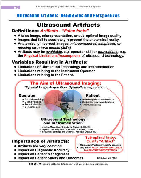
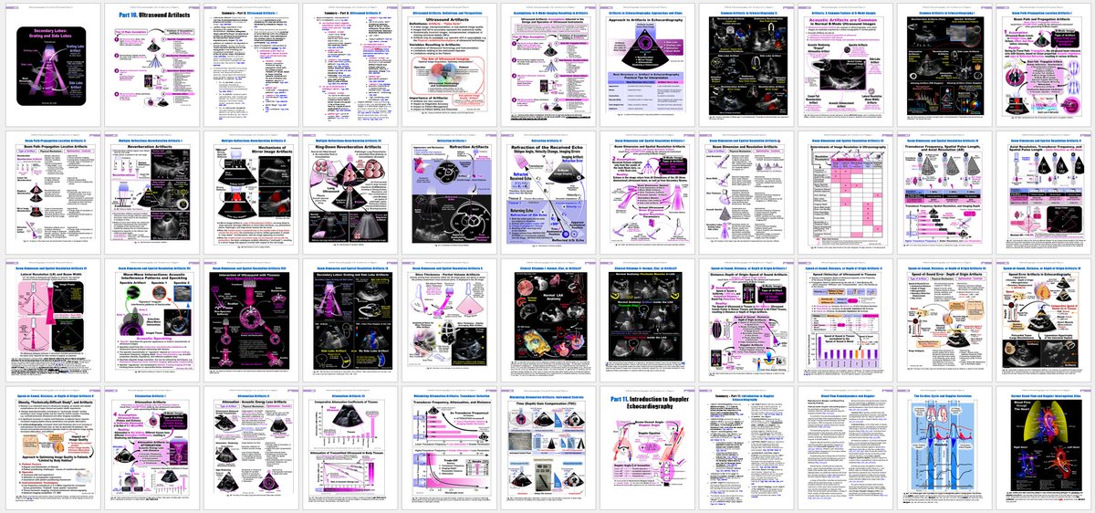
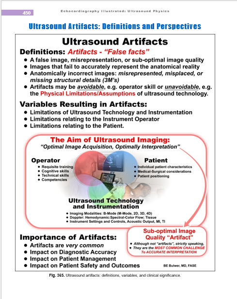
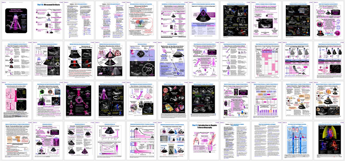
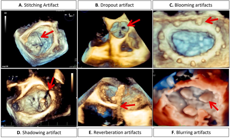
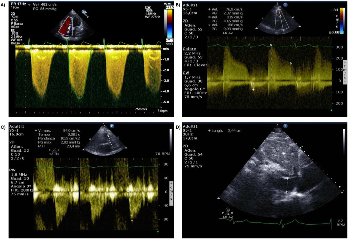
This document provides a uniform & structured approach to managing ultrasound artifacts, including the appearance of the artifact, the mechanism behind the artifact generation, the clinical impact of the artifact on the diagnosis, & examples of real cases. bit.ly/4tKDSvf