Bryan E. Tan, MD

2.3K posts

Bryan E. Tan, MD banner
Bryan E. Tan, MD

Bryan E. Tan, MD

@BryanTanMD

@UChicago Incoming Advanced Cardiac Imaging Fellow | @BCMHeart Cardiology Fellow PGY6 🫀| ACC NCDR LAAO R&P committee | RGH IM 🩺 | Husband, Girl Dad | 🇲🇾🇺🇸

Houston, TX Katılım Nisan 2013
508 Takip Edilen1.1K Takipçiler
Sabitlenmiş Tweet
Bryan E. Tan, MD
Bryan E. Tan, MD@BryanTanMD·
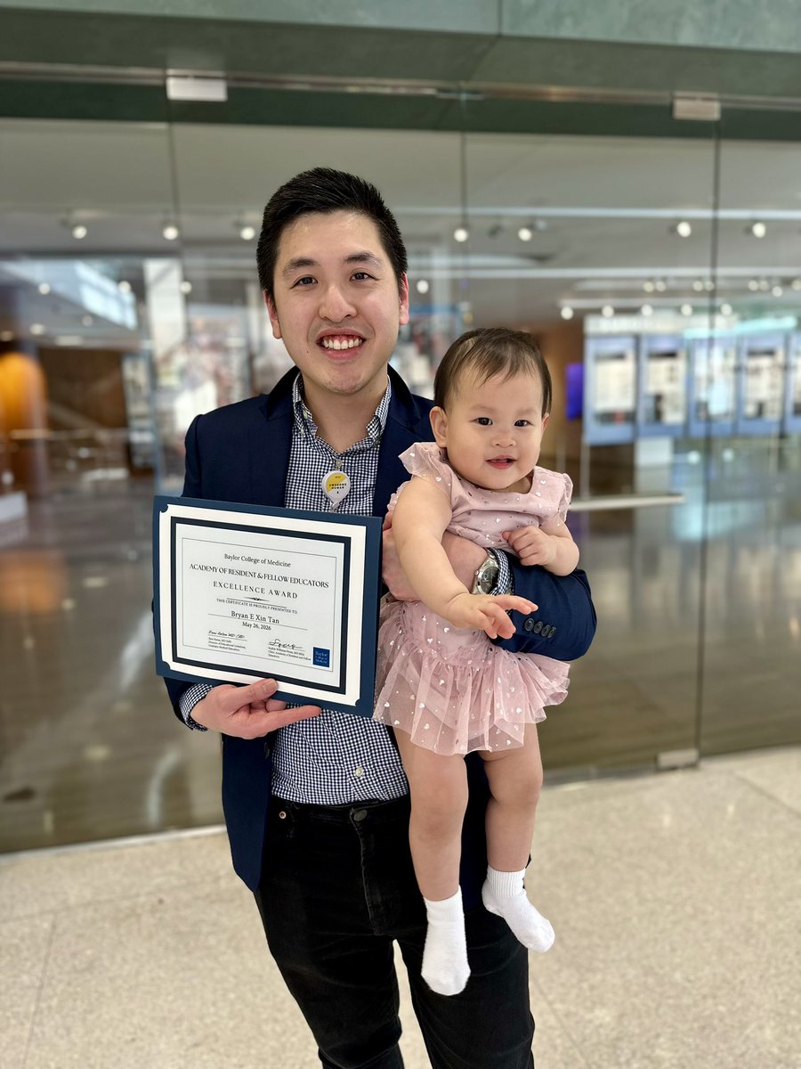
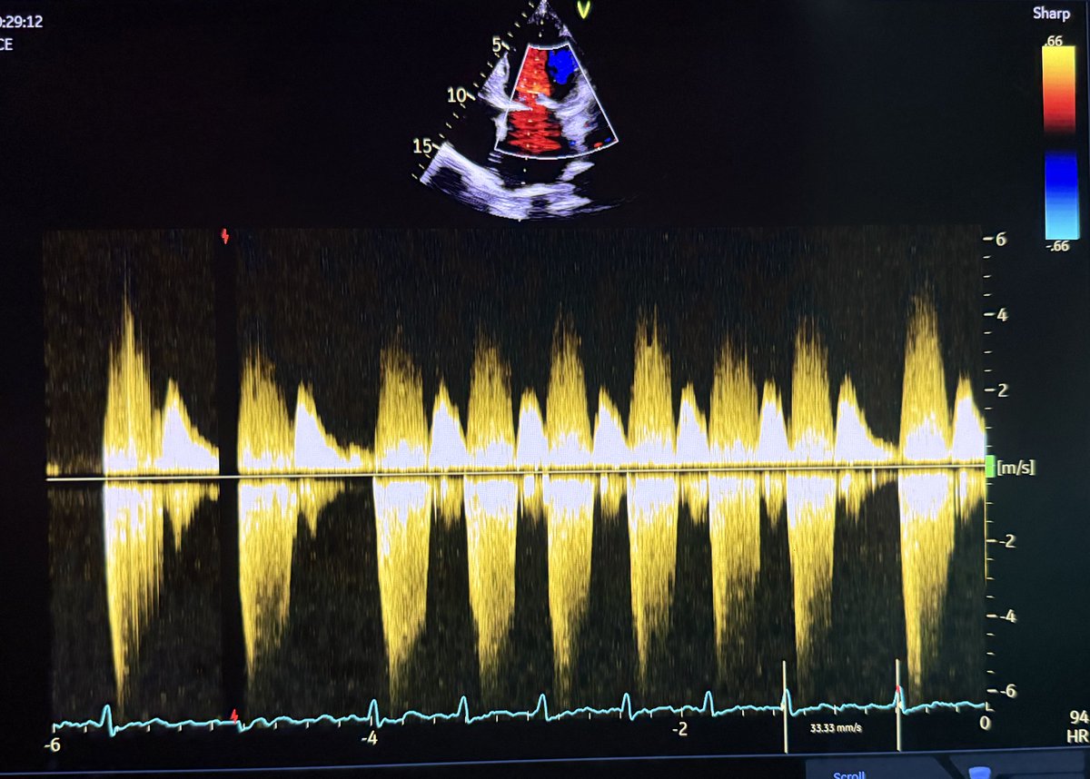
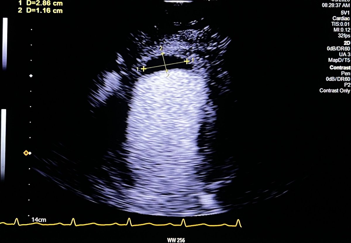
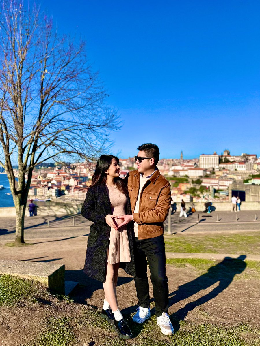
2025 has been an amazing year for our family! Some highlights: ✨Had a blast in Portugal 🇵🇹 for baby moon 🌙 ✨The arrival of our 1st baby 👶🏼 ✨ Matched at 1st choice for Advanced Imaging Fellowship 🩻 ✨Aced Echo boards (95 percentile) Looking forward to another great year!
Bryan E. Tan, MD tweet mediaBryan E. Tan, MD tweet mediaBryan E. Tan, MD tweet mediaBryan E. Tan, MD tweet media
English
5
3
130
16.4K
Bryan E. Tan, MD
Bryan E. Tan, MD@BryanTanMD·
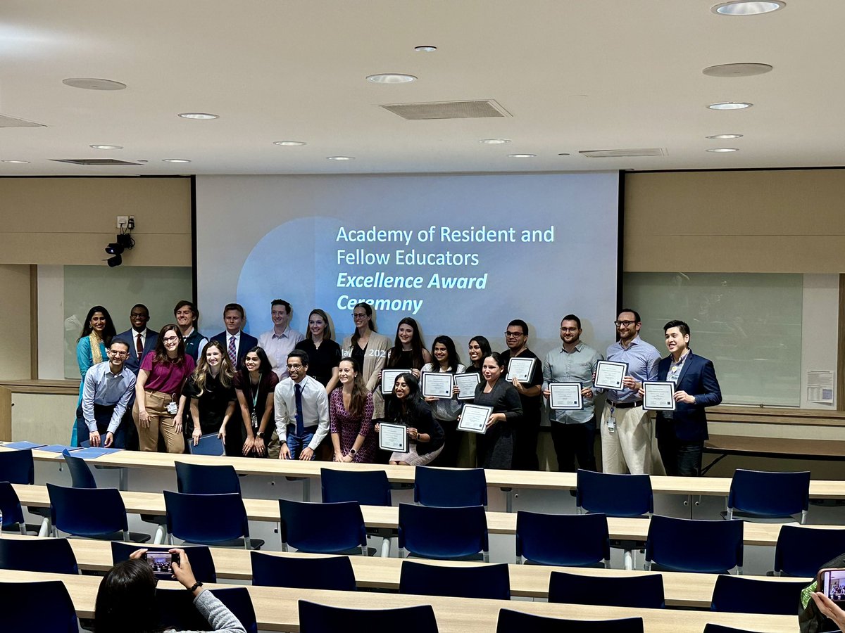
Honored to received the Trainee Excellence Award 🏅from the Academy of Resident and Fellow Educators at @bcmhouston. I’m thankful to my wife & family, program director @arunima_misra and my mentors at @BCMHeart for their unwavering support in my career and in life 😇 #ACCFIT
Bryan E. Tan, MD tweet mediaBryan E. Tan, MD tweet mediaBryan E. Tan, MD tweet mediaBryan E. Tan, MD tweet media
English
3
2
32
2.3K
Bryan E. Tan, MD
Bryan E. Tan, MD@BryanTanMD·
Thank you, Dr. Goyal! That was an incredible talk. Really enjoyed the discussion about Ikigai and growth mindset!
Amit Goyal MD MAS@AmitGoyalMD

It was a privilege to share ideas with this extraordinary group of @bcmhouston fellows about "Finding Meaning, Fulfillment, and Financial Freedom in Medicine". We discussed Ikigai, personal finance, joyfulness, the wheel of life, and so much more. Greateful to @DrHaloot & @arunima_misra for the invitation.

English
1
0
1
582
Bryan E. Tan, MD retweetledi
Mushtaq Bilal, PhD
Mushtaq Bilal, PhD@MushtaqBilalPhD·
Academic publisher Elsevier's profit margin compared to Apple, Google, and Microsoft Apple: 28% Google: 25% Microsoft: 34% Elsevier: 37% with a revenue of $3.9 billion. Elsevier's payment to academic authors and reviewers: $0
English
64
928
3.5K
198.2K
Abdul Mannan Khan Minhas
Abdul Mannan Khan Minhas@AbdulMannan5465·
Published in BMC Cardiovascular Disorders: Burden of cardiovascular disease in Pakistan - insight from global burden of disease study 2019 rdcu.be/ffqkW
Abdul Mannan Khan Minhas tweet media
English
2
7
39
2.9K
Bryan E. Tan, MD retweetledi
Benigno Valderrábano Salas
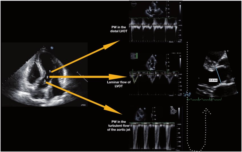
🫀 Did you know where the recommendation to place the pulsed wave Doppler sample volume 0.5–1 cm from the aortic valve to measure LVOT VTI comes from? The answer is more interesting than it seems. It doesn’t come from a single study or an experiment designed for that purpose. It comes from a historical chain spanning nearly 40 years: 🔬 1982–1984 — The physical foundation Pasipoularides and Murgo demonstrated using invasive catheters and mathematical models that in aortic stenosis there is a real zone of flow acceleration in the LVOT, just proximal to the valve, without any second anatomic obstruction. Pure hemodynamics — no Doppler yet. 👉 Bird et al. Circulation 1982 → doi.org/10.1161/01.CIR… 👉 Pasipoularides et al. Am J Physiol 1984 → doi.org/10.1152/ajphea… 📐 1984 — The apical 5-chamber view Lewis, Kuo and Quinones were the first to validate cardiac output measurement using pulsed wave Doppler from the cardiac apex. They described placing the sample volume “immediately proximal to the aortic valve leaflets” — but without specifying any distance in centimeters. 👉 Lewis et al. Circulation 1984 → doi.org/10.1161/01.CIR… 📏 1985 — The first numerical distance Skjaerpe, Hegrenaes and Hatle (the Norwegian group) were the first to quantify this in Doppler: they empirically observed that flow acceleration began 0.5 to 1.5 cm proximal to the valve, and placed the sample volume just proximal to that zone. They directly cited Pasipoularides as supporting evidence. This was the first time a numerical distance appeared in the technique. 👉 Skjaerpe et al. Circulation 1985 → doi.org/10.1161/01.CIR… 📊 1986–1988 — Practical consolidation Otto et al. used ~1.0 cm. Oh, Tajik and the Mayo Clinic group explicitly established the range of 0.5 to 1.0 cm in 100 patients, justifying it as necessary to avoid the subvalvular acceleration zone. This is the figure we all recognize today. 👉 Otto et al. JACC 1986 → doi.org/10.1016/S0735-… 👉 Zoghbi et al. Circulation 1986 → doi.org/10.1161/01.CIR… 👉 Oh et al. JACC 1988 → doi.org/10.1016/0735-1… 📋 2002 — It becomes “official” Quinones, Otto, Zoghbi and colleagues codified it in the ASE guidelines as “~5 mm proximal to the aortic valve”… but without citing any specific study to support it. It had already become expert consensus. 👉 Quiñones et al. JASE 2002 → doi.org/10.1067/mje.20… ⚔️ 2017 — The debate reopens Baumgartner et al. (EACVI/ASE) maintained the 0.5–1 cm recommendation. However, Hahn and Pibarot responded with a critical letter pointing out that the original articles from the 1980s measured at the aortic annulus, not 0.5–1 cm below it, and that moving away from the annulus introduces errors due to the elliptical and irregular shape of the subannular LVOT. 👉 Baumgartner et al. Eur Heart J Cardiovasc Imaging 2017 → doi.org/10.1093/ehjci/… 👉 Hahn & Pibarot. JASE 2017 → doi.org/10.1016/j.echo… 💡 Bottom line: The 0.5–1 cm figure was never experimentally validated as the optimal distance. It emerged from empirical observations in the 1980s aimed at avoiding a flow acceleration zone that had been demonstrated with invasive catheters. It was adopted through accumulated clinical practice and later elevated to a formal recommendation by consensus. The debate over whether to measure at the annulus or 0.5–1 cm below it remains open to this day. One of those recommendations we all follow but few know where it actually came from 🙂 Dr Benigno Valderrábano Salas @MDBeni @JaeKOh2 @ottoecho @WilliamZoghbi @ASE360 @EACVIPresident @NephroP @iamritu @PPibarot @hahn_rt @MAecocardio @SISIACOficial @SONECOM_AC @VazyurVasquez @Cardiotweets83 @HEARTof_echo @echobasics
Benigno Valderrábano Salas tweet media
English
17
132
412
54.1K
Bryan E. Tan, MD retweetledi
Jose Ramos Vivas
Jose Ramos Vivas@joseramosvivas·
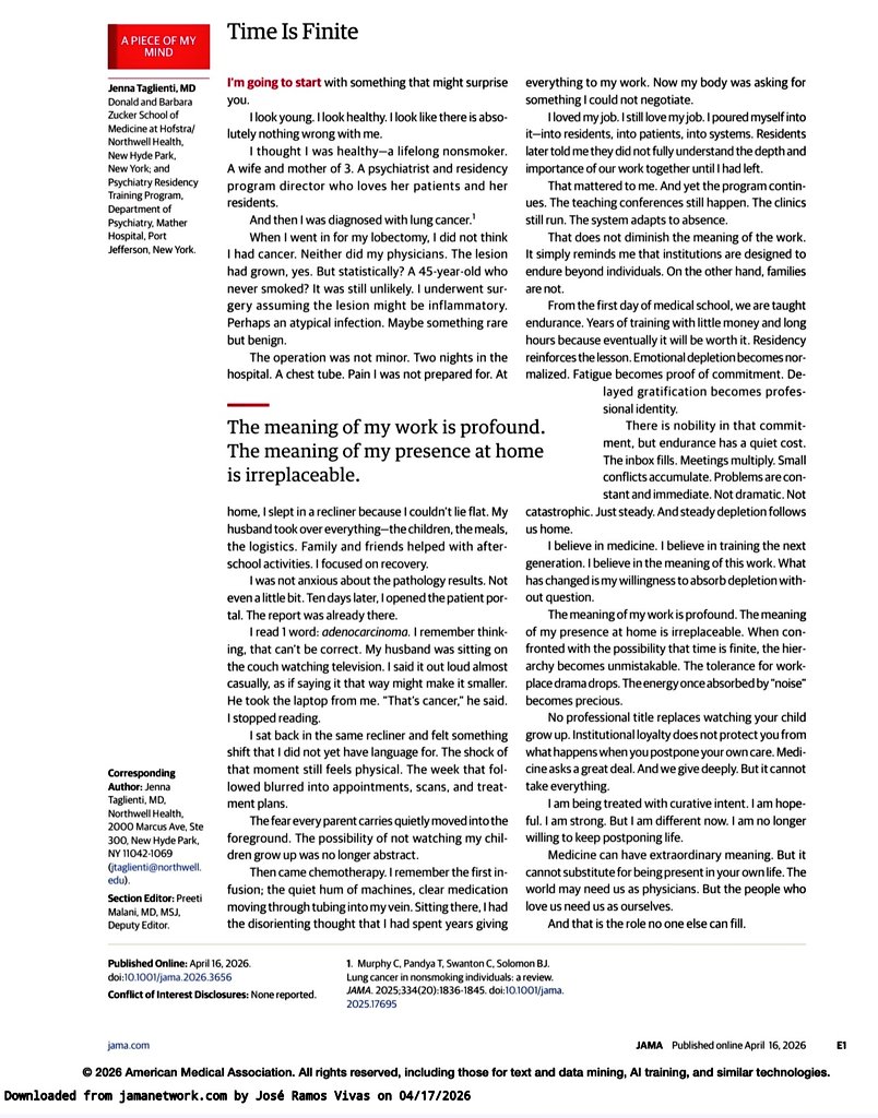
Especial médicos🩺⚕️🏥👨🏻‍⚕️👩🏻‍⚕️. Os dejo esta carta en JAMA. Se la pondré a mis alumnos en la próxima clase: He elegido estas frases : 👇🏻⏰ «La medicina puede tener un significado extraordinario. Pero no puede sustituir el estar presente en tu propia vida. El mundo puede necesitarnos como médicos. Pero las personas que nos aman nos necesitan como nosotros mismos. Y ese es el rol que nadie más puede llenar.» «La residencia refuerza la lección de que las instituciones están diseñadas para perdurar más allá de los individuos. En cambio, las familias no.» «Creo en formar a la próxima generación. Creo en el significado de este trabajo. Lo que ha cambiado es mi disposición a absorber el desgaste sin cuestionarlo.» «Ya no estoy dispuesta a seguir posponiendo la vida. La medicina exige mucho. Y nosotros damos profundamente. Pero no puede tomarlo todo.» «El significado de mi trabajo es profundo. El significado de mi presencia en casa es irremplazable.»
Jose Ramos Vivas tweet media
Español
42
1.7K
3.8K
215.6K
Bryan E. Tan, MD retweetledi
CardiovascularCorner
CardiovascularCorner@TrackYourHeart·
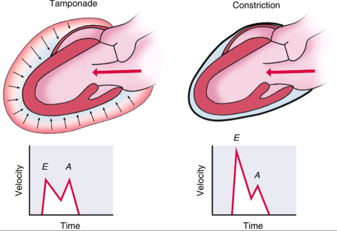
Pericardial Tamponade vs Constrictive Pericarditis ➡️Spot the Difference Tamponade ⬇️ Accumulation of fluid in the pericardial space ⬇️ ↑ intrapericardial pressure ⬇️ impairs diastolic filling throughout the cardiac cycle (early and late diastole). Doppler shows blunted E and A waves due to restricted filling. Constrictive Pericarditis ⬇️ Thickened, non-compliant pericardium ⬇️ rapid early diastolic filling that abruptly halts once volume limit is hit. Doppler shows prominent E wave, reduced A wave, classic for constriction. Recognizing the echocardiographic filling patterns is essential for diagnosis. Ref: Catherine M. Otto, Textbook of Clinical Echocardiography
CardiovascularCorner tweet media
English
1
69
235
11.8K
Chittur Sivaram MD MACP MACC
Chittur Sivaram MD MACP MACC@CASivaram1·
#echofirst ✅ Ultrasound Enhancing Agents (UEA) are indicated for LVO (endocardial definition) & evaluation of cardiac masses ✅ An example ⬇️ where UEA showed unambiguous LV apical & apical septal mural thrombus, not seen on regular 2DE ✅ Thrombus seen ⬇️ in orthogonal views, the plane between endocardium & mass sharply delineated, reduced LVEF ✅ High value yield of UEA & also pretty images @iamritu @argulian @TWeickert78 @PWesslyMD @alex1708ander @DrRajeshG1 @BryanTanMD @AJamilTajik @VLSorrellImages @echo_stepbystep
Chittur Sivaram MD MACP MACC tweet media
English
3
13
50
3.1K